4.2 Digestion and Absorption
Digestion
Now, let’s take a trip through the digestive tract to see what happens to that peanut butter sandwich you ate for lunch.
Mouth
Both physical and chemical digestion begin in the mouth or oral cavity which is the point of entry of food into the digestive system. The sandwich is broken into smaller particles by mastication, the chewing action of the teeth. Chemically, your sandwich is still the same, it’s just in smaller pieces. All mammals have teeth and can chew their food to begin the process of physically breaking it down into smaller particles.
The chemical process of digestion begins during chewing as your sandwich mixes with saliva produced by the salivary glands. Saliva contains mucus that moistens food and buffers the pH of the food. Saliva also contains lysozyme which has antibacterial action. It also contains an enzyme called salivary amylase that begins the process of digesting the starches (a type of carbohydrate) in your meal. Food does not spend enough time in the mouth to allow all the carbohydrates to break down, but salivary amylase continues acting until it is inactivated by stomach acids. The chewing and wetting action provided by the teeth and saliva prepare the food into a mass called the bolus for swallowing. At this point, your sandwich has been in the GI tract for 15-30 seconds and is about 3 inches into its journey. We are ready to swallow the food so we can move on to the next organ in this trip.
The tongue helps in swallowing by moving the bolus from the mouth into the pharynx. The pharynx opens to two passageways: the esophagus and the trachea. The esophagus leads to the stomach and the trachea leads to the lungs. The epiglottis is a flap of tissue that covers the tracheal opening during swallowing to direct the bolus down the esophagus and prevent food from entering the lungs.
Esophagus
The esophagus is a muscular tube that connects the pharynx to the stomach. It is approximately 10 inches in length and the chewed and softened food takes 5-10 seconds to pass through the esophagus after being swallowed. The smooth muscles of the esophagus undergo peristalsis that pushes the food toward the stomach. The peristaltic wave is unidirectional – it moves food from the mouth toward the stomach, and reverse movement is not possible, except in the case of the vomit reflex. The peristaltic movement of the esophagus is an involuntary reflex; it takes place in response to the act of swallowing.
Sphincters are muscles that surround tubes and serve as valves, closing the tube when the sphincters contract and opening it when they relax. Food passes from the esophagus into the stomach at the lower esophageal sphincter (also called the gastroesophageal or cardiac sphincter). The lower esophageal sphincter relaxes to let food pass into the stomach, and then contracts to prevent stomach acids from backing up into the esophagus. When the lower esophageal sphincter does not completely close, the stomach’s contents can move back up into the esophagus, causing heartburn or gastroesophageal reflux disease (GERD). This movement of the stomach contents back into the esophagus is called reflux.
Stomach
The stomach is a muscular sac composed of three layers of muscle. These muscles relax and contract to produce a powerful churning motion. The stomach is only approximately 6 inches long but can expand to hold between 4-6 cups of food. Once your chewed and softened sandwich arrives in the stomach, stomach cells secrete gastric juices. Gastric juices consist of water, hydrochloric acid (HCl), mucus, and enzymes. HCl is a strong acid that lowers the pH of the stomach and has two functions related to digestion. First, HCl kills bacteria and other microorganisms that may be in the food. Second, HCl begins the digestion of protein by activating an enzyme that digests proteins in the stomach. Stomach walls are made of muscle proteins, so why is it that the enzymes do not digest the cells of the stomach? The stomach has specialized cells that secrete a thick layer of mucus. This mucus forms a thick lining that protects the stomach walls from HCl and enzymes. If the mucus breaks down, then the HCl and enzymes can come in contact with the stomach lining and cause sores or ulcers to form. The enzymes found in gastric juices begin the digestion of some proteins and fats. However, there is minimal digestion and absorption in the stomach.
In addition to this chemical digestion, the stomach also participates in mechanical digestion. Within a few moments after food enters your stomach, the muscles surrounding the stomach begin to contract in a wave like manner. These muscular contractions are a unique type of peristalsis that mixes and softens the food with gastric juices to create a semisolid liquid called chyme. It is fair to say that long before your sandwich exits through the pyloric sphincter after 2-6 hours, it bears little resemblance to the sandwich you ate. Different types of food take different amounts of time to process. Foods heavy in carbohydrates empty fastest, followed by protein rich foods. Meals with a high fat content remain in the stomach the longest.
Its numerous digestive functions notwithstanding, there is only one stomach function necessary to life: the production of intrinsic factor. The intestinal absorption of vitamin B12, which is necessary for both the production of mature red blood cells and normal neurological functioning, cannot occur without intrinsic factor. People who undergo total gastrectomy (stomach removal) – for life-threatening stomach cancer, for example – can survive with minimal digestive dysfunction if they receive vitamin B12 injections.
Small intestine
Chyme released from the stomach passes through the pylorus or pyloric sphincter and enters the small intestine, which is the primary digestive organ in the body. At this point, the sandwich you ate has been turned into chyme, the starches in the bread and proteins in the peanut butter are partially digested. There has been a lot of mechanical digestion but the fats in the peanut butter are largely undigested and none of your nutrients have been absorbed yet.
Immediately upon arrival in the small intestine, the pancreas secretes bicarbonate to neutralize stomach acid and digestive enzymes to chemically digest all three macronutrients. When there is fat in the small intestine, the gallbladder secretes bile to aid in fat digestion. The small intestine has three sections; the duodenum, jejunum, and ileum. Most chemical digestion occurs in the duodenum and is facilitated by the pancreatic enzymes. However, the small intestine’s absorptive cells in the jejunum and ileum also synthesize digestive enzymes to finish digesting carbohydrates, fats, and proteins and prepare them for absorption. In addition to peristalsis, another type of mechanical action called segmentation occurs in the small intestine. Segmentation involves contraction of ring-like intestinal muscles which helps to move the chyme in a back and forth, or “sloshing” motion. This helps to mix the enzymes in with the chyme and enhances absorption of nutrients.
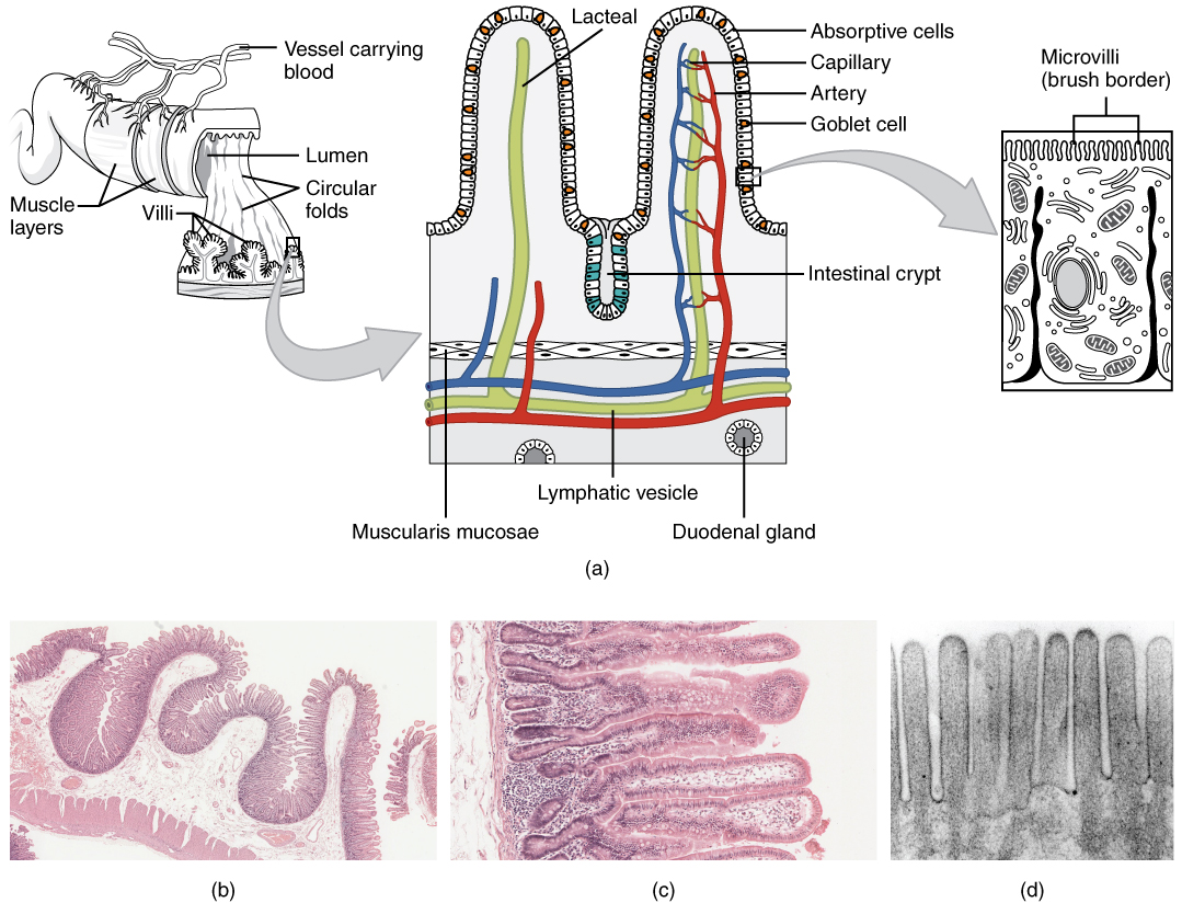
Not only is the small intestine where most chemical digestion occurs, it is also where almost all absorption occurs. The longest part of the GI tract, the small intestine, is between 13-20 feet long in a living person (but about twice as long in a cadaver due to the loss of muscle tone). Since this makes it about five times longer than the large intestine, you might wonder why it is called “small.” In fact, its name derives from its relatively smaller diameter of only about 1 inch, compared with 3 inches for the large intestine. As we’ll see shortly, in addition to its length, the folds and projections of the lining of the small intestine work to give it an enormous surface area, which is approximately 200 m2, more than 100 times the surface area of your skin. This large surface area is necessary for complex processes of digestion and absorption that occur within it. The small intestine has a very unique anatomical design. The interior is designed to maximize surface area and increase our capacity for absorption. The interior wall of the small intestine is highly folded and the folds are lined with small finger-like projections called villi. Running up into every villus is a capillary and lymphatic vessel. Each villus also has an outer layer of epithelial cells called absorptive cells or enterocytes. Each of these cells is lined with even smaller projections called microvilli. The area of the microvilli is referred to as the brush border and it is here that the final steps of digestion occur before nutrients are absorbed.
Figure 4.3 Anatomy of the Small Intestine

By the time your sandwich has finished passing through the small intestine, you have completely broken down the sandwich into individual carbohydrates, fats, and proteins. These nutrients were absorbed into your enterocytes. The leftover mixture is still very watery because as your meal made its way through the GI tract, liquids were secreted into the GI tract in almost every organ. Overall, your sandwich spent about 3-6 hours in the small intestine. In general, high carbohydrate foods are digested and absorbed fastest and fatty foods take the longest to digest and absorb. However, the type of carbohydrate and size of the meal can impact transit time too.
Large intestine (Colon)
The residue of chyme that enters the large intestine, also known as the colon, contains few nutrients except water, which is reabsorbed as the residue lingers in the large intestine, typically for 12 to 24 hours. Thus, it may not surprise you that the large intestine can be completely removed without significantly affecting digestive functioning. For example, in severe cases of inflammatory bowel disease, the large intestine can be removed by a procedure known as a colectomy. Often, a new fecal pouch can be crafted from the small intestine and sutured to the anus, but if not, an ileostomy can be created by bringing the distal ileum (lower part of the small intestine) through the abdominal wall, allowing the watery chyme to be collected in a bag-like adhesive appliance.
The large intestine is also home to your gut microbiome, discussed in more detail in the next section. Bacteria that reside in the colon can produce some vitamins and short chain fatty acids which can be absorbed into the bloodstream. Vitamin K is one of the vitamins produced by your intestinal bacteria which makes it very difficult to study. Even if dietary vitamin K intake is controlled, it can be produced by bacteria in the gut. This is also why babies often receive a shot of vitamin K after birth, before bacteria has had a chance to take up residence in their guts.
Absorption
Once the food is digested into individual nutrients, the nutrients are then absorbed across the brush border to enter either the capillaries or lymphatic vessels. Water soluble nutrients such as sugars, amino acids, and water soluble vitamins and minerals, enter tiny blood vessels known as capillaries and are transported to the liver via the hepatic portal vein. Once in the liver, some nutrients are stored and some released into circulation. Because the capillaries are tiny blood vessels and the blood is primarily composed of water, only water soluble nutrients can be transported from the gut to the liver via the hepatic portal vein. Any nutrients that are not water soluble, must be packaged as a chylomicron in the enterocytes before they can enter the blood. Chylomicrons will be discussed in more detail in chapter 6. These chylomicrons are too large to fit in the capillaries so must be transported via lymph. Fat soluble nutrients enter the lymph vessels which bypass the liver and are deposited directly into the bloodstream. Nutrients transported via lymph include fats and the fat soluble vitamins A, D, E, and K. Lymph vessels also contain white blood cells that aid your immune system.
Media Attributions
- Histology of the Small Intestines © OpenStax College is licensed under a CC BY (Attribution) license
