15 Forensic Anthropology
Ashley Kendell, Ph.D., California State University, Chico
Alex Perrone, M.A., M.S.N, R.N., P.H.N., Butte Community College
Colleen Milligan, Ph.D., California State University, Chico
This chapter is a revision from “Chapter 15: Bioarchaeology and Forensic Anthropology” by Ashley Kendell, Alex Peronne, and Colleen Milligan. In Explorations: An Open Invitation to Biological Anthropology, first edition, edited by Beth Shook, Katie Nelson, Kelsie Aguilera, and Lara Braff, which is licensed under CC BY-NC 4.0.
Content Warning and Disclaimer: This chapter includes images of human remains as well as discussions centered on human skeletal analyses. All images are derived from casts, sketches, nonhuman skeletal material, as well as non-Indigenous skeletal materials curated within the CSU, Chico Human Identification Lab, and the Hartnett-Fulginiti donated skeletal collection.
Learning Objectives
- Define forensic anthropology as a subfield of biological anthropology.
- Describe the seven steps carried out during skeletal analysis.
- Outline the four major components of the biological profile.
- Contrast the four categories of trauma.
- Explain how to identify the different taphonomic agents that alter bone.
- Discuss ethical considerations for forensic anthropology.
Forensic anthropology is a subfield of biological anthropology and an applied area of anthropology. Forensic anthropologists use skeletal analysis to gain information about humans in the present or recent past, then they apply this information within a medicolegal context. This means that forensic anthropologists specifically conduct their analysis on recently deceased individuals (typically within the last 50 years) as part of investigations by law enforcement. Forensic anthropologists can assist law enforcement agencies in several different ways, including aiding in the identification of human remains whether they are complete, fragmentary, burned, scattered, or decomposed. Additionally, forensic anthropologists can help determine what happened to the deceased at or around the time of death as well as what processes acted on the body after death (e.g., whether the remains were scattered by animals, whether they were buried in the ground, or whether they remained on the surface as the soft tissue decomposed).
Many times, because of their expertise in identifying human skeletal remains, forensic anthropologists are called to help with outdoor search-and-recovery efforts, such as locating remains scattered across the surface or carefully excavating and documenting buried remains. In other cases, forensic anthropologists recover remains after natural disasters or accidents, such as fire scenes, and can help identify whether each bone belongs to a human or an animal. Forensic anthropology spans a wide scope of contexts involving the law, including incidences of mass disasters, genocide, and war crimes.
A point that can be somewhat confusing for students is that although the term forensic is included in this subfield of biological anthropology, there are many forensic techniques that are not included in the subfield. Almost exclusively, forensic anthropology deals with skeletal analysis. While this can include the comparison of antemortem (before death) and postmortem (after death) radiographs to identify whether remains belong to a specific person, or using photographic superimposition of the cranium, it does not include analyses beyond the skeleton. For example, blood-spatter analysis, DNA analysis, fingerprints, and material evidence collection do not fall under the scope of forensic anthropology.
So, what can forensic anthropologists glean from bones alone? Forensic anthropologists can address a number of questions about a human individual based on their skeletal remains. Some of those questions are as follows: How old was the person? Was the person biologically male or female? How tall was the person? What happened to the person at or around their time of death? Were they sick? The information from the skeletal analysis can then be matched with missing persons records, medical records, or dental records, aiding law enforcement agencies with identifications and investigations.
Skeletal Analysis
Forensic anthropology relies on skeletal analysis to reveal information about the deceased. The methodology and approaches outlined below are specific to the United States. Forensic anthropological methods differ depending on the country conducting an investigation. In the United States, there are typically seven steps or questions to the process:
- Is it bone?
- Is it human?
- Is it modern or archaeological?
- How many individuals are present or what is the minimum number of individuals (MNI)?
- Who is it?
- Is there evidence of trauma before or around the time of death?
- What happened to the remains after death?
Is It Bone?
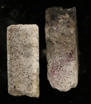
One of the most important steps in any skeletal analysis starts with determining whether or not material suspected to be bone is in fact bone. Though it goes without saying that a forensic anthropologist would only carry out analysis on bone, this step is not always straightforward. Whole bones are relatively easy to identify, but determining whether or not something is bone becomes more challenging once it becomes fragmentary. As an example, in high heat such as that seen on fire scenes, bone can break into pieces. During a house fire with fatalities, firefighters watered down the burning home. After the fire was extinguished, the sheetrock (used to construct the walls of the home) was drenched and crumbled. The crumbled sheetrock was similar in color and form to burned, fragmented bone, therefore mistakable for human remains (Figure 15.1). Forensic anthropologists on scene were able to separate the bones from the construction material, helping to confirm the presence of bone and hence the presence of individual victims of the fire. In this case, forensic anthropologists were able to recognize the anatomical and layered structure of bone and were able to distinguish it from the uniform and unlayered structure of sheetrock.
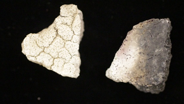
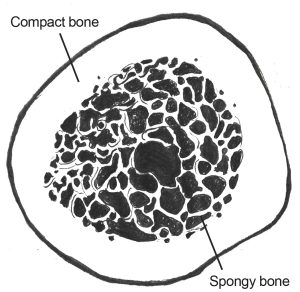
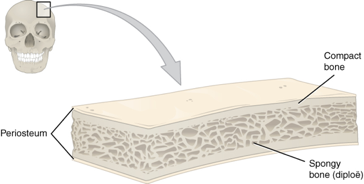
As demonstrated by the example above, both the macrostructure (visible with the naked eye) and microstructure (visible with a microscope) of bone are helpful in bone identification. Bones are organs in the body made up of connective tissue. The connective tissue is hardened by a mineral deposition, which is why bone is rigid in comparison to other connective tissues such as cartilage (Tersigni-Tarrant and Langley 2017, 82–83; White and Folkens 2005, 31). In a living body, the mineralized tissue does not make up the only component of bone—there are also blood, bone marrow, cartilage, and other types of tissues. However, in dry bone, two distinct layers of the bone are the most helpful for identification. The outer layer is made up of densely arranged osseous (bone) tissue called compact (cortical) bone. The inner layer is composed of much more loosely organized, porous bone tissue whose appearance resembles that of a sponge, hence the name spongy (trabecular) bone. Knowing that most bone contains both layers helps with the macroscopic identification of bone (Figures 15.2, 15.3). For example, a piece of coconut shell might look a lot like a fragment of a human skull bone. However, closer inspection will demonstrate that coconut shell only has one very dense layer, while bone has both the compact and spongy layers.
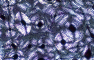
The microscopic identification of bone relies on knowledge of osteons, or bone cells (Figure 15.4). Under magnification, bone cells are visible in the outer, compact layer of bone. The bone cells are arranged in a concentric pattern around blood vessels for blood supply. The specific shape of the cells can help differentiate, for example, a small piece of PVC (white plastic) pipe from a human bone fragment (Figure 15.5).
Is It Human?
Once it has been determined that an object is bone, the next logical step is to identify whether the bone belongs to a human or an animal. Forensic anthropologists are faced with this question in everyday practice because human versus nonhuman bone identification is one of the most frequent requests they receive from law enforcement agencies.
There are many different ways to distinguish human versus nonhuman bone. The morphology (the shape/form) of human bone is a good place for students to start. Identifying the 206 bones in the adult human skeleton and each bone’s distinguishing features (muscle attachment sites, openings and grooves for nerves and blood vessels, etc.) is fundamental to skeletal analysis.
Nevertheless, there are many animal bones and human bones that look similar. For example, the declawed skeleton of a bear paw looks a lot like a human hand, pig molars appear similar to human molars, and some smaller animal bones might be mistaken for those of an infant. To add to the confusion, fragmentary bone may be even more difficult to identify as human or nonhuman. However, several major differences between human and nonhuman vertebrate bone help distinguish the two.
Forensic anthropologists pay special attention to the density of the outer, compact layer of bone in both the cranium and in the long bones. Human cranial bone has three distinctive layers. The spongy bone is sandwiched between the outer (ectocranial) and inner (endocranial) compact layers. In most other mammals, the distinction between the spongy and compact layers is not always so definite. Secondly, the compact layer in nonhuman mammal long bones can be much thicker than observed in human bone. Due to the increased density of the compact layer, nonhuman bone tends to be heavier than human bone (Figure 15.6).
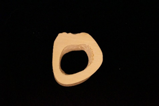
The size of a bone can also help determine whether it belongs to a human. Adult human bones are larger than subadult or infant bones. However, another major difference between human adult bones and those of a young individual or infant human can be attributed to development and growth of the epiphyses (ends of the bone). The epiphyses of human subadult bones are not fused to the shaft (Figure 15.7). Therefore, if a bone is small and it is suspected to belong to a human subadult or infant, the epiphyses would not be fused. Many small animal bones appear very similar in form compared to adult human bones, but they are much too small to belong to an adult human. Yet they can be eliminated as subadult or infant bones if the epiphyses are fused to the shaft.
Is It Modern or Archaeological?
Forensic anthropologists work with modern cases that fall within the scope of law enforcement investigations. Accordingly, it is important to determine whether discovered human remains are archaeological or forensic in nature. Human remains that are historic are considered archeaological. The scientific study of human remains from archaeological sites is called bioarchaeology.
Dig Deeper: Bioarchaeology
For readers who are interested in the sister subfield of bioarchaeology, which studies human remains and material culture from the past, please refer to chapter 8 of Bioarchaeology: Interpreting Human Behavior from Skeletal Remains, in TRACES: An Open Invitation to Archaeology (Blatt, Michael, and Bright forthcoming).
A forensic anthropologist should begin their analysis by reviewing the context in which the remains were discovered. This will help them understand a great deal about the remains, including determining whether they are archaeological or forensic in nature as well as considering legal and ethical issues associated with the collection, analysis, and storage of human remains (see “Ethics and Human Rights” section of this chapter for more information).
The “context” refers to the relationship the remains have to the immediate area in which they were found. This includes the specific place where the remains were found, the soil or other organic matter immediately surrounding the remains, and any other objects or artifacts in close proximity to the body. For example, imagine that a set of remains has been located during a house renovation. The remains are discovered below the foundation. Do the remains belong to a murder victim? Or was the house built on top of an ancient burial ground? Observing information from the surroundings can help determine whether the remains are archaeological or modern. How long ago was the foundation of the house erected? Are there artifacts in close proximity to the body, such as clothing or stone tools? These are questions about the surroundings that will help determine the relative age of the remains.
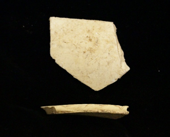
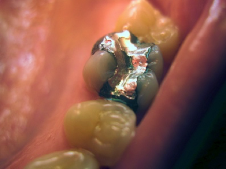
Clues directly from the skeleton may also indicate whether the remains are archaeological or modern. For example, tooth fillings can suggest that the individual was alive recently (Figure 15.8). In fact, filling material has changed over the decades, so the specific type of material used to fix a cavity can be matched with specific time periods. Gold was used in dental work in the past, but more recently composite (a mixture of plastic and fine glass) fillings have become more common.
How Many Individuals Are Present?
What Is MNI?
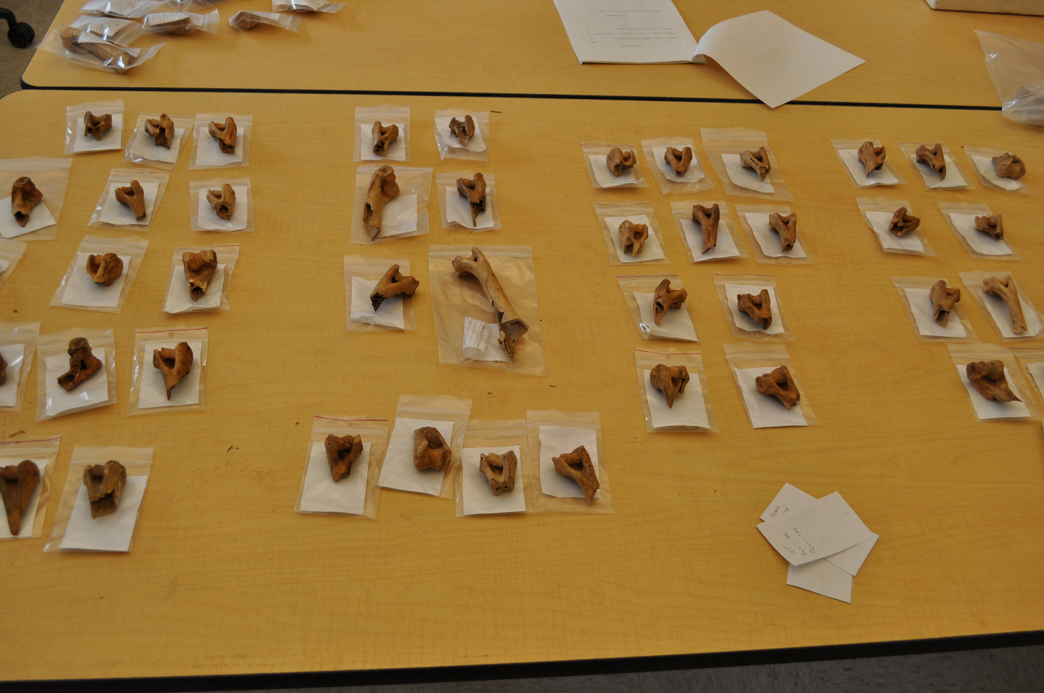
Another assessment that an anthropologist can perform is the calculation of the number of individuals in a mixed burial assemblage. Because not all burials consist of a single individual, it is important to burial assemblage be able to estimate the number of individuals in a forensic context. Quantification of the number of individuals in a burial assemblage can be done through the application of a number of methods, including the following: the Minimum Number of Individuals (MNI), the Most Likely Number of Individuals (MLNI), and the Lincoln Index (LI). The most commonly used method in biological anthropology, and the focus of this section, is determination of the MNI.
The MNI presents “the minimum estimate for the number of individuals that contributed to the sample” (Adams and Konigsberg 2008, 243). Many methods of calculating MNI were originally developed within the field of zooarchaeology for use on calculating the number of individuals in faunal or animal assemblages (Adams and Konigsberg 2008, 241). What MNI calculations provide is a lowest possible count for the total number of individuals contributing to a skeletal assemblage. Traditional methods of calculating MNI include separating a skeletal assemblage into categories according to the individual bone and the side the bone comes from and then taking the highest count per category and assigning that as the minimum number (Figure 15.9).
Why Calculate MNI?
In a forensic context, the determination of MNI is most applicable in cases of mass graves, commingled burials, and mass fatality incidents. The term commingled is applied to any burial assemblage in which individual skeletons are not separated into separate burials. As an example, the authors of this chapter have observed commingling of remains resulting from mass fatality wildfire events. Commingled remains may also be encountered in events such as a plane or vehicle crash. It is important to remember that in any forensic context, MNI should be referenced and an MNI of one should be substantiated by the fact that there was no repetition of elements associated with the case.
Constructing the Biological Profile
Who Is It?
“Who is it?” is one of the first questions that law enforcement officers ask when they are faced with a set of skeletal remains. To answer this question, forensic anthropologists construct a biological profile (White and Folkens 2005, 405). A biological profile is an individual’s identifying characteristics, or biological information, which include the following: biological sex, age at death, stature, population affinity, skeletal variation, and evidence of trauma and pathology.
Forensic anthropologists typically construct a biological profile to help positively identify a deceased person. The following section will lay out each component of the biological profile and briefly review standard methodology used for each.
Assessing Biological Sex
Assessment of biological sex is often one of the first things considered when establishing a biological profile because several other parts, such as age and stature estimations, rely on an assessment of biological sex to make the calculations more accurate.
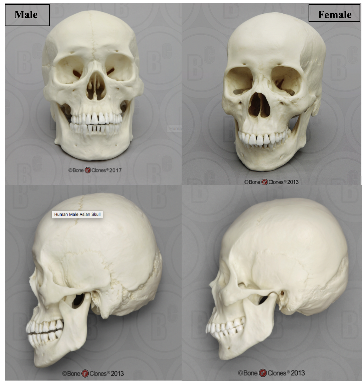
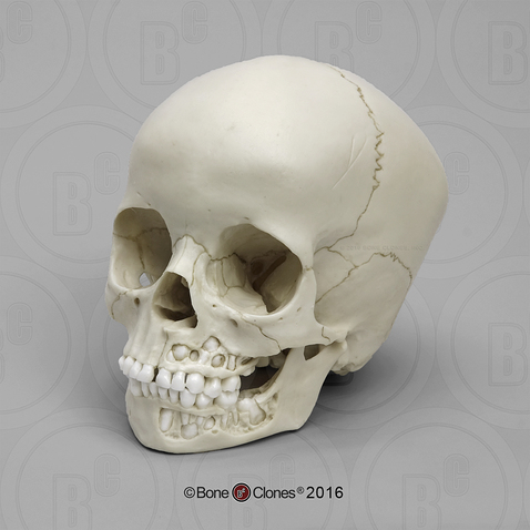
Assessment of biological sex focuses on differences in both morphological (form or structure) and metric (measured) traits in individuals. When assessing morphological traits, the skull and the pelvis are the most commonly referenced areas of the skeleton. These differences are related to sexual dimorphism usually varying in the amount of robusticity seen between males and females. Robusticity deals with strength and size; it is frequently used as a term to describe a large size or thickness. In general, males will show a greater degree of robusticity than females. For example, the length and width of the mastoid process, a bony projection located behind the opening for the ear, is typically larger in males. The mastoid process is an attachment point for muscles of the neck, and this bony projection tends to be wider and longer in males. In general, cranial features tend to be more robust in males (Figure 15.10).
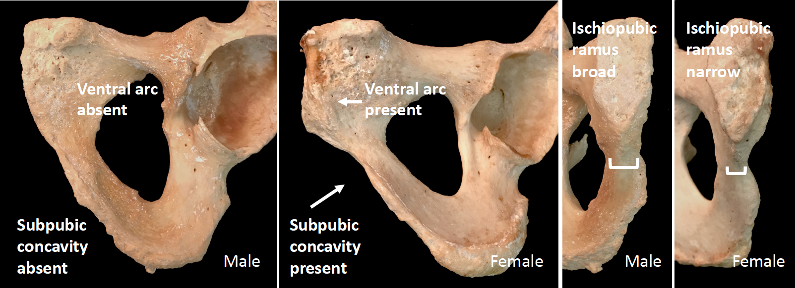
When considering the pelvis, the features associated with the ability to give birth help distinguish females from males. During puberty, estrogen causes a widening of the female pelvis to allow for the passage of a baby. Several studies have identified specific features or bony landmarks associated with the widening of the hips, and this section will discuss one such method. The Phenice Method (Phenice 1969) is traditionally the most common reference used to assess morphological characteristics associated with sex. The Phenice Method specifically looks at the presence or absence of (1) a ventral arc, (2) the presence or absence of a subpubic concavity, and (3) the width of the medial aspect of the ischiopubic ramus (Figure 15.11). When present, the ventral arc, a ridge of bone located on the ventral surface of the pubic bone, is indicative of female remains. Likewise the presence of a subpubic concavity and a narrow medial aspect of the ischiopubic ramus is associated with a female sex estimation. Assessments of these features, as well as those of the skull (when both the pelvis and skull are present), are combined for an overall estimation of sex.
Metric analyses are also used in the estimation of sex. Measurements taken from every region of the body can contribute to estimating sex through statistical approaches that assign a predictive value of sex. These approaches can include multiple measurements from several skeletal elements in what is called multivariate (multiple variables) statistics. Other approaches consider a single measurement, such as the diameter of the head of the femur, of a specific element in a univariate (single variable) analysis (Berg 2017, 152–156).
It is important to note that, although forensic anthropologists usually begin assessment of biological profile with biological sex, there is one major instance in which this is not appropriate. The case of two individuals found in California, on July 8, 1979, is one example that demonstrates the effect age has on the estimation of sex. The identities of the two individuals were unknown; therefore, law enforcement sent them to a lab for identification. A skeletal analysis determined that the remains represented one adolescent male and one adolescent female, both younger than 18 years of age. This information did not match with any known missing children at the time.
In 2015, the cold case was reanalyzed, and DNA samples were extracted. The results indicated that the remains were actually those of two girls who went missing in 1978. The girls were 15 years old and 14 years old at the time of death. It is clear that the 1979 results were incorrect, but this mistake also provides the opportunity to discuss the limitations of assessing sex from a subadult skeleton.
Assessing sex from the human skeleton is based on biological and genetic traits associated with females and males. These traits are linked to differences in sexual dimorphism and reproductive characteristics between females and males. The link to reproductive characteristics means that most indicators of biological sex do not fully manifest in prepubescent individuals, making estimations of sex unreliable in younger individuals (SWGANTH 2010b). This was the case in the example of the 14-year-old girl. When examined in 1979, her remains were misidentified as male because she had not yet fully developed female pelvic traits.
Sex vs. Gender
Biological sex is a different concept than gender. While biological anthropologists can estimate sex from the skeleton, estimating an individual’s gender would require a greater context because gender is defined culturally rather than biologically. Take, for example, an individual who identifies as transgender. This individual has a gender identity that is different from their biological sex. The gender identity of any individual depends on factors related to self-identification, situation or context, and cultural factors. While in the U.S. we have historically thought of sex and gender as binary concepts (male or female), many cultures throughout the world recognize several possible gender identities. In this sense, gender is seen as a continuous or fluid variable rather than a fixed one.
Historically, forensic anthropologists have used a binary construct to categorize human skeletal remains as either male or female (with the accompanying categories of probable male, probable female, and indeterminate). In the case of transgender and gender nonconforming individuals, the binary approach to sex assessment may delay or hinder identification efforts (Buchanan 2014; Schall, Rogers, and Deschamps-Braly 2020; Tallman, Kincer, and Plemons 2021). As such, many forensic anthropologists have begun to address the inherent problems associated with a binary approach to sex identification and to explore ways of assessing social identity and self-identified gender using skeletal remains and forensic context.
For the duration of this section, the term transgender refers to individuals whose gender identity differs from the sex assigned at birth (Schall, Rogers, and Deschamps-Braly 2020:2). Transgender individuals transition from one gender binary to another, such as male-to-female (MTF) or female-to-male (FTM). While many of the gender-affirming procedures available to trans and gender-nonconforming individuals are focused on soft tissue modifications (e.g., breast augmentation, genital reconstruction, hormone therapies, etc.), there are a number of gender-affirmation surgeries that do leave a permanent record on the skeleton. Generally speaking, FTM transgender people are reported to undergo fewer surgical procedures than do MTF transgender people (Buchanan 2014). The discussion below focuses on Facial Feminization Surgery (FFS), which leaves a permanent record on the human skeleton that may be used to help make an identification.
FFS refers to a combination of procedures focused on sexually dimorphic features of the face, with the intent of transforming typically male facial features into more feminine forms. Facial Feminization Surgery procedures were developed by Dr. Douglas Ousterhout, a San Francisco based cranio-maxillofacial surgeon, in the mid-1980s (Schall, Rogers, and Deschamps-Braly 2020:2). FFS can include one or a combination of the following: hairline lowering, forehead reduction and contouring, brow lift, reduction rhinoplasty, cheek enhancement, lift lift, lip filling, chin contouring, jaw contouring, and/or tracheal shave (Buchanan 2014; Schall, Rogers, and Deschamps-Braly 2020:2). Of the procedures outlined previously, four are known to directly affect the facial skeleton: forehead contouring, rhinoplasty, chin contouring, and jaw contouring (Buchanan 2014; Schall, Rogers, and Deschamps-Braly 2020:2).
Because FFS procedures have been widely documented in the medical (and more recently the forensic anthropological) literature, there are a number of indicators that a forensic anthropologist can use to make more informed evaluations of gender, including evidence of bone remodeling in sexually dimorphic regions of the skull (e.g., forehead, chin, jawline), as well as the presence of plates, pins, or other surgical hardware that may be evidence of FFS (Buchanan 2014; Schall, Rogers, and Deschamps-Braly 2020; Tallman, Kincer, and Plemons 2021). Additionally, some forensic anthropologists suggest cautiously integrating contextual information from the scene, such as personal effects, material evidence, and recovery scene information, into their evaluation of an individual’s social identity (Beatrice and Soler 2016; Birkby, Fenton, and Anderson 2008; Soler and Beatrice 2018; Soler et al. 2019; Tallman, Kincer, and Plemons 2021; Winburn, Schoff, and Warren 2016). The ultimate goal of many skeletal analyses is to make a positive identification on a set of unidentified remains.
Assessment of Population Affinity
Population affinity is another component of the biological profile. We use the term population affinity to refer to the variation seen among modern populations—variation that is both genetic and environmentally driven. The word affinity refers to similarities or relationships between individuals. As forensic anthropologists, we compare an unknown individual to multiple reference groups and look for the degree of similarity in observable traits with those groups. As noted previously, population affinity can aid law enforcement in their identification of missing persons or unknown skeletal remains.
Within the field of anthropology, the estimation of population affinity has a contentious history, and early attempts at classification were largely based on the erroneous assumption that an individual’s phenotype (outward appearance) was correlated with their innate intelligence and abilities (see Chapter 13 for a more in-depth discussion of the history of the race concept). The use of the term race is deeply embedded in the social context of the United States. In any other organism/living thing, groups divided according to the biological race concept would be defined as a separate subspecies. The major issue with applying the biological race concept to humans is that there are not enough differences between any two populations to separate on a genetic basis. In other words, biological races do not exist in human populations. However, the concept of race has been perpetuated and upheld by sociocultural constructs of race.
The conundrum for forensic anthropologists is the fact that while races do not exist on a biological level, we still socially recognize and categorize individuals based on their phenotype. Clearly, our phenotype is an important factor in not only how we are viewed by others but also how we identify ourselves. It is also a commonly reported variable. Often labeled as “race,” we are asked to report how we self-identify on school applications, government identification, surveys, census reports, and so forth. It follows then that when a person is reported missing, the information commonly collected by law enforcement and sometimes entered into a missing person’s database includes their age, biological sex, stature, and “race.” Therefore, the more information a forensic anthropologist can provide regarding the individual’s physical characteristics, the more he or she can help to narrow the search.
As an exercise, create a list of all of the women you know who are between the ages of 18 and 24 and approximately 5’ 4” to 5’ 9” tall. You probably have several dozen people on the list. Now, consider how many females you know who are between the ages of 18 and 24, are approximately 5’ 4” to 5’ 9” tall, and are Vietnamese. Your list is going to be significantly shorter. That’s how missing persons searches go as well. The more information you can provide regarding a decedent’s phenotype, the fewer possible matches law enforcement are left to investigate. This is why population affinity has historically been included as a part of the biological profile.
In an effort to combat the erroneous assumptions tied to the race concept, forensic anthropologists have attempted to reframe this component of the biological profile. The term race is no longer used in casework and teaching. Historically, the word ancestry is and was deemed a more appropriate way to describe an individual’s phenotype. However, in more recent years, forensic anthropologists have begun using the term population affinity, recognizing that we are basing our analysis on the similarities we see based on the reference samples we have available (Winburn and Algee-Hewitt 2021). An important note here is that it is possible to hinder identifications and harm individuals when tools like estimations of population affinity are misapplied, misinterpreted, or misused. For this reason, the field of forensic anthropology has ongoing conversations about the appropriateness of this analysis in the biological profile (Bethard and DiGangi 2020; Stull et al. 2021).
Traditionally, population affinity was accomplished through a visual inspection of morphological variants of the skull (morphoscopics). These methods focused on elements of the facial skeleton, including the nose, eyes, and cheek bones. However, in an effort to reduce subjectivity, nonmetric cranial traits are now assessed within a statistical framework to help anthropologists better interpret their distribution among living populations (Hefner and Linde 2018). Based on the observable traits, a macromorphoscopic analysis will allow the practitioner to create a statistical prediction of geographic origin. In essence, forensic anthropologists are using human variation in the estimation of geographic origin, by referencing documented frequencies of nonmetric skeletal indicators or macromorphoscopic traits.
Population affinity is also assessed through metric analyses. The computer program Fordisc is an anthropological tool used to estimate different components of the biological profile, including ancestry, sex, and stature. When using Fordisc, skeletal measurements are input into the computer software, and the program employs multivariate statistical classification methods, including discriminant function analysis, to generate a statistical prediction for the geographic origin of unknown remains based on the comparison of the unknown to the reference samples in the software program. Fordisc also calculates the likelihood of the prediction being correct, as well as how typical the metric data is for the assigned group.
Estimating Age-at-Death
Estimating age-at-death from the skeleton relies on the measurement of two basic physiological processes: (1) growth and development and (2) degeneration (or aging). From fetal development on, our bones and teeth grow and change at a predictable rate. This provides for relatively accurate age estimates. After our bones and teeth cease to grow and develop, they begin to undergo structural changes, or degeneration, associated with aging. This does not happen at such predictable rates and, therefore, results in less accurate or larger age-range estimations.
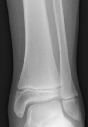
During growth and development stages, two primary methods used for estimations of age of subadults (those under the age of 18) are epiphyseal union and dental development. Epiphyseal union (or epiphyseal fusion) refers to the appearance and closure of the epiphyseal plates between the primary centers of growth in a bone and the subsequent centers of growth (see Figure 15.7). Prior to complete union, the cartilaginous area between the primary and secondary centers of growth is also referred to as the growth plates (Schaefer, Black, and Scheuer 2009). Different areas of the skeleton have documented differences in the appearance and closure of epiphyses, making this a reliable method for aging subadult remains (SWGANTH 2013).
As an example of its utility in the identification process, epiphyseal development was used to identify two subadult victims of a fatal fire in Flint, Michigan, in February 2010. The remains represented two young girls, ages three and four. Due to the intensity of the fire, the subadult victims were differentiated from each other through the appearance of the patella, the kneecap. The patella is a bone that develops within the tendon of the quadriceps muscle at the knee joint. The patella begins to form around three to four years of age (Cunningham, Scheuer, and Black 2016, 407–409). In the example above, radiographs of the knees showed the presence of a patella in the four-year-old girl and the absence of a clearly discernible patella in the three-year-old.
Dental development begins during fetal stages of growth and continues until the complete formation and eruption of the adult third molars (if present). The first set of teeth to appear are called deciduous or baby teeth. Individuals develop a total of 20 deciduous teeth, including incisors, canines, and molars. These are generally replaced by adult dentition as an individual grows (Figure 15.12). A total of 32 teeth are represented in the adult dental arcade, including incisors, canines, premolars, and molars. When dental development is used for age estimations, researchers use both tooth-formation patterns and eruption schedules as determining evidence. For example, the crown of the tooth forms first followed by the formation of the tooth root. During development, an individual can exhibit a partially formed crown or a complete crown with a partially formed root. The teeth generally begin the eruption process once the crown of the tooth is complete. The developmental stages of dentition are one of the most reliable and consistent aging methods for subadults (Langley, Gooding, and Tersigni-Tarrant 2017, 176–177).
Degenerative changes in the skeleton typically begin after 18 years of age, with more prominent changes developing after an individual reaches middle adulthood (commonly defined as after 35 years of age in osteology). These changes are most easily seen around joint surfaces of the pelvis, the cranial vault, and the ribs. In this chapter, we focus on the pubic symphysis surfaces of the pelvis and the sternal ends of the ribs, which show metamorphic changes from young adulthood to older adulthood. The pubic symphysis is a joint that unites the left and right halves of the pelvis. The surface of the pubic symphysis changes during adulthood, beginning as a surface with pronounced ridges (called billowing) and flattening with a more distinct rim to the pubic symphysis as an individual ages. As with all metamorphic age changes, older adults tend to develop lipping around the joint surfaces as well as a breakdown of the joint surfaces. The most commonly used method for aging adult skeletons from the pubic symphysis is the Suchey-Brooks method (Brooks and Suchey 1990; Katz and Suchey 1986). This method divides the changes seen with the pubic symphysis into six phases based on macroscopic age-related changes to the surface. Figure 15.13 provides a visual of the degenerative changes that typically occur on the pubic symphysis.
The sternal end of the ribs, the anterior end of the rib that connects via cartilage to the sternum, is also used in age estimations of adults. This method, first developed by M. Y. İşcan and colleagues, considers both the change in shape of the sternal end as well as the quality of the bone (İşcan, Loth, and Wright 1984; İşcan, Loth, and Wright 1985). The sternal end first develops a billowing appearance in young adulthood. The bone typically develops a wider and deeper cupped end as an individual ages. Older adults tend to exhibit bony extensions of the sternal end rim as attaching cartilage ossifies. Figure 15.14 provides a visual of the degenerative changes that typically occur in sternal rib ends.
Estimating Stature
Stature, or height, is one of the most prominently recorded components of the biological profile. Our height is recorded from infancy through adulthood. Doctor’s appointments, driver’s license applications, and sports rosters all typically involve a measure of stature for an individual. As such, it is also a component of the biological profile nearly every individual will have on record. Bioarchaeologists and forensic anthropologists use stature estimation methods to provide a range within which an individual’s biological height would fall. Biological height is a person’s true anatomical height. However, the range created through these estimations is often compared to reported stature, which is typically self-reported and based on an approximation of an individual’s true height (Ousley 1995).
In June 2015, two men were shot and killed in Granite Bay, California, in a double homicide. Investigators were able to locate surveillance camera footage from a gas station where the two victims were spotted in a car with another individual believed to be the perpetrator in the case. The suspect, sitting behind the victims in the car, hung his right arm out of the window as the car drove away. The search for the perpetrator was eventually narrowed down to two suspects. One suspect was 5’ 8” while the other suspect was 6’ 4”, representing almost a foot difference in height reported stature between the two. Forensic anthropologists were given the dimensions of the car (for proportionality of the arm) and were asked to calculate the stature of the suspect in the car from measurements of the suspect’s forearm hanging from the window. Approximate lengths of the bones of the forearm were established from the video footage and used to create a predicted stature range. Stature estimations from skeletal remains typically look at the correlation between the measurements of any individual bone and the overall measurement of body height. In the case above, the length of the right forearm pointed to the taller of the two suspects who was subsequently arrested for the homicide.
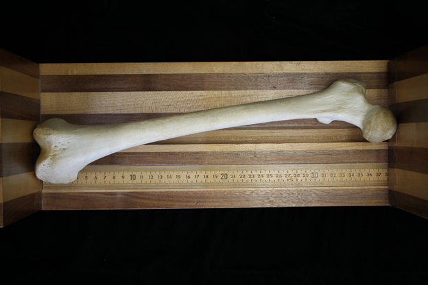
Certain bones, such as the long bones of the leg, contribute more to our overall height than others and can be used with mathematical equations known as regression equations. Regression methods examine the relationship between variables such as height and bone length and use the correlation between the variables to create a prediction interval (or range) for estimated stature. This method for calculating stature is the most commonly used method (SWGANTH 2012). Figure 15.15 shows the measurement of the bicondylar length of the femur for stature estimations.
Identification Using Individualizing Characteristics
One of the most frequently requested analyses within the forensic anthropology laboratory is assistance with the identification of unidentified remains. While all components of a biological profile, as discussed above, can assist law enforcement officers and medical examiners to narrow down the list of potential identifications, a biological profile will not lead to a positive identification. The term positive identification refers to a scientifically validated method of identifying previously unidentified remains. Presumptive identifications, however, are not scientifically validated; rather, they are based on circumstances or scene context. For example, if a decedent is found in a locked home with no evidence of forced entry but the body is no longer visually identifiable, it may be presumed that the remains belong to the homeowner. Hence, a presumptive identification.
The medicolegal system ultimately requires that a positive identification be made in such circumstances, and a presumptive identification is often a good way to narrow down the pool of possibilities. Biological profile information also assists with making a presumptive identification based on an individual’s phenotype in life (e.g., what they looked like). As an example, a forensic anthropologist may establish the following components of a biological profile: white male, between the ages of 35 and 50, approximately 5’ 7” to 5’ 11.” While this seems like a rather specific description of an individual, you can imagine that this description fits dozens, if not hundreds, of people in an urban area. Therefore, law enforcement can use the biological profile information to narrow their pool of possible identifications to include only white males who fit the age and height outlined above. Once a possible match is found, the decedent can be identified using a method of positive identification.
Positive identifications are based on what we refer to as individualizing traits or characteristics, which are traits that are unique at the individual level. For example, brown hair is not an individualizing trait as brown is the most common hair color in the U.S. But, a specific pattern of dental restorations or surgical implants can be individualizing, because it is unlikely that you will have an exact match on either of these traits when comparing two individuals.
A number of positive methods are available to forensic anthropologists, and for the remainder of this section we will discuss the following methods: comparative medical and dental radiography and identification of surgical implants.
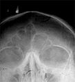
Comparative medical and dental radiography is used to find consistency of traits when comparing antemortem records (medical and dental records taken during life) with images taken postmortem (after death). Comparative medical radiography focuses primarily on features associated with the skeletal system, including trabecular pattern (internal structure of bone that is honeycomb in appearance), bone shape or cortical density (compact outer layer of bone), and evidence of past trauma, skeletal pathology, or skeletal anomalies. Other individualizing traits include the shape of various bones or their features, such as the frontal sinuses (Figure 15.16).
Comparative dental radiography focuses on the number, shape, location, and orientation of dentition and dental restorations in antemortem and postmortem images. While there is not a minimum number of matching traits that need to be identified for an identification to be made, the antemortem and postmortem records should have enough skeletal or dental consistencies to conclude that the records did in fact come from the same individual (SWGANTH 2010a). Consideration should also be given to population-level frequencies of specific skeletal and dental traits. If a trait is particularly common within a given population, it may not be a good trait to utilize for positive identification.
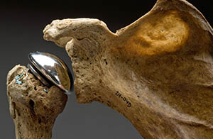
Surgical implants or devices can also be used for identification purposes (Figure 15.17). These implements are sometimes recovered with human remains. One of the ways forensic anthropologists can use surgical implants to assist in decedent identification is by providing a thorough analysis of the implant and noting any identifying information such as serial numbers, manufacturer symbols, and so forth. This information can then sometimes be tracked directly to the manufacturer or the place of surgical intervention, which may be used to identify unknown remains (SWGANTH 2010a).
Special Topic: Trans Doe Task Force
The Trans Doe Task Force (TDTF) is a Trans-led nonprofit organization that investigates cases involving LGBTQ+ missing and murdered persons. The organization specifically focuses on transgender and gender-variant cases, providing connections between law enforcement agencies, medical examiner offices, forensic anthropologists, and forensic genetic genealogists to increase the chances of identification. Additionally, the TDTF curates a data repository of missing, murdered, and unclaimed LGBTQ+ individuals, and they continuously try innovative approaches to identify these individuals, whose lived gender identity may not match their biological sex.
For more information visit transdoetaskforce.org
Trauma Analysis
Types of Trauma
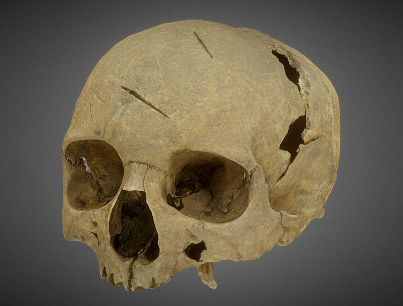
Within the field of anthropology, trauma is defined as an injury to living tissue caused by an extrinsic force or mechanism (Lovell 1997:139). Forensic anthropologists can assist a forensic pathologist by providing an interpretation of the course of events that led to skeletal trauma. Typically, traumatic injury to bone is classified into one of four categories, defined by the trauma mechanism. A trauma mechanism refers to the force that produced the skeletal modification and can be classified as (1) sharp force, (2) blunt force, (3) projectile, or (4) thermal (burning). Each type of trauma, and the characteristic pattern(s) associated with that particular categorization, will be discussed below.
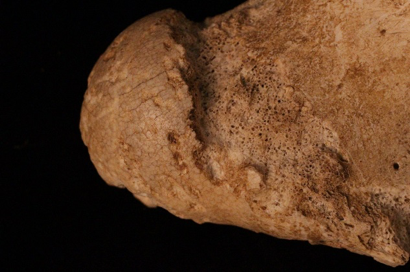
First, let’s consider sharp-force trauma, which is caused by a tool that is edged, pointed, or beveled—for example, a knife, saw, or machete (SWGANTH 2011). The patterns of injury resulting from sharp-force trauma include linear incisions created by a sharp, straight edge; punctures; and chop marks (Figure 15.18; SWGANTH 2011). When observed under a microscope, an anthropologist can often determine what kind of tool created the bone trauma. For example, a power saw cut will be discernible from a manual saw cut.
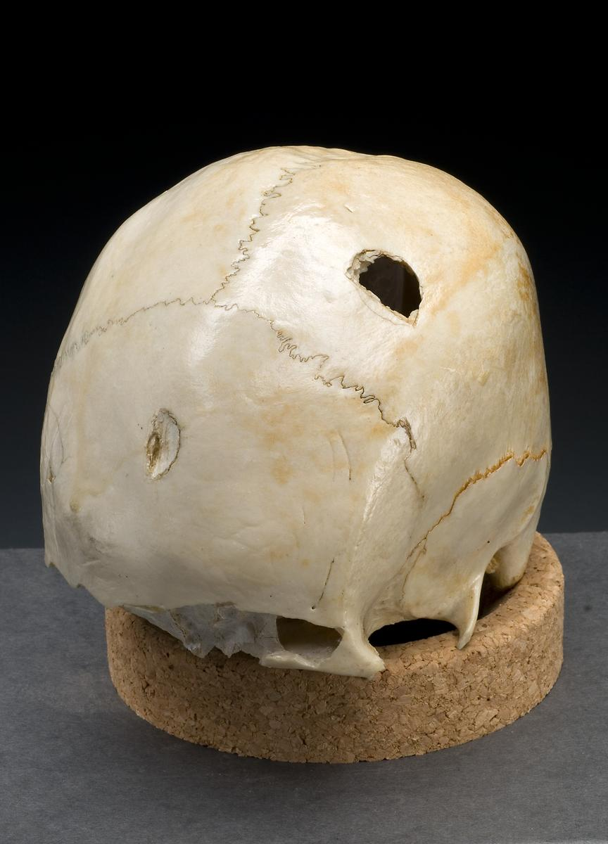
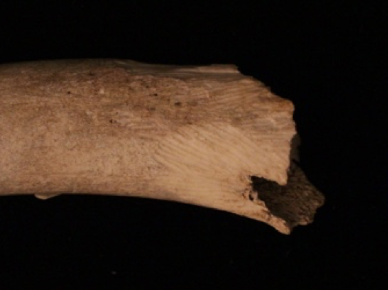
Second, blunt-force trauma is defined as “a relatively low-velocity impact over a relatively large surface area” (Galloway 1999, 5). Blunt-force injuries can result from impacts from clubs, sticks, fists, and so forth. Blunt-force impacts typically leave an injury at the point of impact but can also lead to bending and deformation in other regions of the bone. Depressions, fractures, and deformation at and around the site of impact are all characteristics of blunt-force trauma (Figure 15.19). As with sharp-force trauma, an anthropologist attempts to interpret blunt-force injuries, providing information pertaining to the type of tool used, the direction of impact, the sequence of impacts, if more than one, and the amount of force applied.
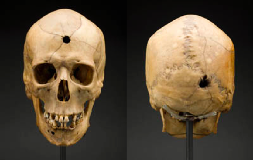
Third, projectile trauma refers to high-velocity trauma, typically affecting a small surface area (Galloway 1999, 6). Projectile trauma results from fast-moving objects such as bullets or shrapnel. It is typically characterized by penetrating defects or embedded materials (Figure 15.20). When interpreting injuries resulting from projectile trauma, an anthropologist can often offer information pertaining to the type of weapon used (e.g., rifle vs. handgun), relative size of the bullet (but not the caliber of the bullet), the direction the projectile was traveling, and the sequence of injuries if there are multiple present.
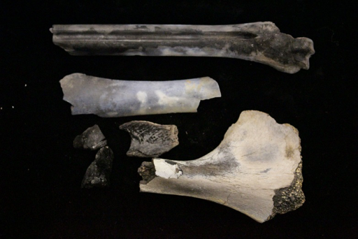
Finally, thermal trauma is a bone alteration that results from bone exposure to extreme heat. Thermal trauma can result in cases of house or car fires, intentional disposal of a body in cases of homicidal violence, plane crashes, and so on. Thermal trauma is most often characterized by color changes to bone, ranging from yellow to black (charred) or white (calcined). Other bone alterations characteristic of thermal trauma include delamination (flaking or layering due to bone failure), shrinkage, fractures, and heat-specific burn patterning. When interpreting injuries resulting from thermal damage, an anthropologist can differentiate between thermal fractures and fractures that occurred before heat exposure, thereby contributing to the interpretation of burn patterning (e.g., was the individual bound or in a flexed position prior to the fire?).
While there are characteristic patterns associated with the four categories of bone trauma, it is also important to note that these bone alterations do not always occur independently of different trauma types. An individual’s skeleton may present with multiple different types of trauma, such as a projectile wound and thermal trauma. Therefore, it is important that the anthropologist recognize the different types of trauma and interpret them appropriately.
Timing of Injury
Another important component of any anthropological trauma analysis is the determination of the timing of injury (e.g., when did the injury occur). Timing of injury is traditionally split into one of three categories: antemortem (before death), perimortem (at or around the time of death), and postmortem (after death). This classification system differs slightly from the classification system used by the pathologist because it specifically references the qualities of bone tissue and bone response to external forces. Therefore, the perimortem interval (at or around the time of death) means that the bone is still fresh and has what is referred to as a green bone response, which can extend past death by several weeks or even months. For example, in cold or freezing temperatures a body can be preserved for extended periods of time, increasing the perimortem interval, while in desert climates decomposition is accelerated, thereby significantly decreasing the postmortem interval (Galloway 1999, 12). Antemortem injuries (occurring well before death and not related to the death incident) are typically characterized by some level of healing, in the form of a fracture callus or unification of fracture margins. Finally, postmortem injuries (occurring after death, while bone is no longer fresh) are characterized by jagged fracture margins, resulting from a loss of moisture content during the decomposition process (Galloway 1999, 16). In general, all bone traumas should be classified according to the timing of injury, if possible. This information will help the medical examiner or pathologist better understand the circumstances surrounding the decedent’s death, as well as events occurring during life and after the final disposition of the body.
The Role of the Forensic Anthropologist in Trauma Analysis
Within the medicolegal system, forensic anthropologists are often called upon by the medical examiner, forensic pathologist, or coroner to assist with an interpretation of trauma. The forensic anthropologist’s main focus in any trauma analysis is the underlying skeletal system—as well as, sometimes, cartilage. Analysis and interpretation of soft tissue injuries fall within the purview of the medical examiner or pathologist. It is also important to note that the main role of the forensic anthropologist is to provide information pertaining to skeletal injury to assist the medical examiner/pathologist in their final interpretation of injury. Forensic anthropologists do not hypothesize as to the cause of death of an individual. Instead, a forensic anthropologist’s report should include a description of the injury (e.g., trauma mechanism, number of injuries, location, timing of injury); documentation of the injury, which may be utilized in court testimony (e.g., photographs, radiographs, measurements); and, if applicable, a statement as to the condition of the body and state of decomposition, which may be useful for understanding the depositional context (e.g., how long has the body been exposed to the elements; was it moved or in its original location; are any of the alterations to bone due to environmental or faunal exposure instead of intentional human modification).
Taphonomy
What Happened to the Remains After Death?
The majority of the skeletal analysis process revolves around the identity of the deceased individual. However, there is one last, very important question that forensic anthropologists should ask: What happened to the remains after death? Generally speaking, processes that alter the bone after death are referred to as taphonomic changes (refer to Chapter 7 for a discussion regarding taphonomy and the fossil record).
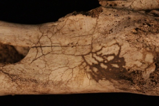
The term taphonomy was originally used to refer to the processes through which organic remains mineralize, also known as fossilization. Within the context of biological anthropology, the term taphonomy is better defined as the study of what happens to human remains after death (Komar and Buikstra 2008). Initial factors affecting a body after death include processes such as decomposition and scavenging by animals. However, taphonomic processes encompass much more than the initial period after death. For example, plant root growth can leach minerals from bone, leaving a distinctive mark. Sunlight can bleach human remains, leaving exposed areas whiter than those that remained buried. Water can wear the surface of the bone until it becomes smooth.
Some taphonomic processes can help a forensic anthropologist estimate the relative amount of time that human remains have been exposed to the elements. For example, root growth through a bone would certainly indicate a body was buried for more than a few days. Forensic anthropologists must be very careful when attempting to estimate time since death based on taphonomic processes because environmental conditions can greatly influence the rate at which taphonomic processes progress. For example, in cold environments, tissue may decay slower than in warm, moist environments.
Forensic anthropologists must contend with taphonomic processes that affect the preservation of bones. For example, high acidity in the soil can break down human bone to the point of crumbling. In addition, when noting trauma, they must be very careful not to confuse postmortem (after death) bone damage with trauma.
|
Taphonomic Process |
Definition |
|---|---|
|
Rodent Gnawing
|
When rodents, such as rats and mice, chew on bone, they leave sets of parallel grooves. The shallow grooves are etched by the rodent’s incisors. |
|
Carnivore Damage
|
Carnivores may leave destructive dental marks on bone. The tooth marks may be visible as pit marks or punctures from the canines, as well as extensive gnawing or chewing of the ends of the bones to retrieve marrow. |
|
Burned Bone
|
Fire causes observable damage to bone. Temperature and the amount of time bone is heated affect the appearance of the bone. Very high temperatures can crack bone and result in white coloration. Color gradients are visible in between high and lower temperatures, with lower temperatures resulting in black coloration from charring. Cracking can also reveal information about the directionality of the burn. |
|
Root Etching
|
Plant roots can etch the outer surface of bone, leaving grooves where the roots attached as they leached nutrients. During this process, the plant’s roots secrete acid that breaks down the surface of the bone.
|
|
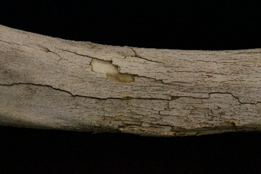
Weathering
|
Many different environmental conditions affect bone. River transport can smooth the surface of the bone due to water abrasion. Sunlight can bleach the exposed surface of bone. Dry and wet environments or the mixture of both types of environments can cause cracking and exfoliation of the surface. Burial in different types of soil can cause discoloration, and exposure can cause degreasing. |
|
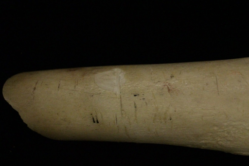
Cut Marks
|
Humans may alter bone by cutting, scraping, or sawing it directly or in the process of removing tissue. The groove pattern—that is, the depth and width of the cuts—can help identify the tool used in the cutting process. |
Ethics and Human Rights
Working with human remains requires a great deal of consideration and respect for the dead. Forensic anthropologists have to think about the ethics of our use of human remains for scientific purposes. How do we conduct casework in the most respectable manner possible? While there are a wide range of ethical considerations to consider when contemplating a career in forensic anthropology, this chapter will focus on two major categories: working with human remains and acting as an expert within the medicolegal system.
Working with Human Remains
Forensic anthropologists work with human remains in a number of contexts, including casework, excavation, research, and teaching. When working with human remains, it is always important to use proper handling techniques. To prevent damage to skeletal remains, bones should be handled over padded surfaces. Skulls should never be picked up by placing fingers in the eye orbits, foramen magnum (hole at the base of the skull for entry of the spinal cord), or through the zygomatic arches (cheekbones). Human remains, whether related to casework, fieldwork, donated skeletal collections, or research, were once living human beings. It is important to always bear in mind that work with remains should be ingrained with respect for the individual and their relatives. In addition to fieldwork, casework, and teaching, anthropologists are often invited to work with remains that come from a bioarchaeological context or from a human rights violation. While this discussion of ethics is not comprehensive, two case examples will be provided below in which an anthropologist must consider the ethical standards outlined above.
Modern Human Rights Violations
Forensic anthropologists may also be called to participate in criminal investigations involving human rights violations. Anthropological investigations may include assistance with identifications, determination of the number of victims, and trauma analyses. In this role, forensic anthropologists play an integral part in promoting human rights, preventing future human rights violations, and providing the evidence necessary to prosecute those responsible for past events. A few ethical considerations for the forensic anthropologist involved in human rights violations include the use of appropriate standards of identification, presenting reliable and unbiased testimony, and maintaining preservation of evidence. For a more comprehensive history of forensic anthropological contributions to human rights violations investigations, see Ubelaker 2018.
Acting as an Expert in the Medicolegal System
In addition to the ethical considerations involved in working with human skeletal remains, forensic anthropologists must abide by ethical standards when they act as experts within the medicolegal system. The role of the forensic anthropologist within the medicolegal system is primarily to provide information to the medical examiner or coroner that will aid in the identification process or determination of cause and manner of death. Forensic anthropologists also may be called to testify in a court of law. In this capacity, forensic anthropologists should always abide by a series of ethical guidelines that pertain to their interpretation, presentation, and preservation of evidence used in criminal investigations. First and foremost, practitioners should never misrepresent their training or education. When appropriate, outside opinions and assistance in casework should be requested (e.g., consulting a radiologist for radiological examinations or odontologist for dental exams). The best interest of the decedent should always take precedence. All casework should be conducted in an unbiased way, and financial compensation should never be accepted as it can act as an incentive to take a biased stance regarding casework. All anthropological findings should be kept confidential, and release of information is best done by the medical examiner or coroner. Finally, while upholding personal ethical standards, forensic anthropologists are also expected to report any perceived ethical violations committed by their peers.
Ethical standards for the field of forensic anthropology are outlined by the Organization of Scientific Area Committees (OSAC) for Forensic Science, administered by the National Institute of Standards and Technology (NIST). OSAC and NIST recently began an initiative to develop standards that would strengthen the practice of forensic science both in the United States and internationally. OSAC’s main objective is to “strengthen the nation’s use of forensic science by facilitating the development of technically sound forensic science standards and by promoting the adoption of those standards by the forensic science community” (NIST n.d.). Additionally, OSAC promotes the establishment of best practices and other guidelines to ensure that forensic science findings and their presentation are reliable and reproducible (NIST 2023).
Special Topic: Native American Graves Protection and Repatriation Act (NAGPRA)
There is a long history in the United States of systematic disenfranchisement of Native American people, including lack of respect for tribal sovereignty. This includes the egregious treatment of Native American human remains. Over several centuries, thousands of Native American remains were removed from tribal lands and held at institutions in the United States, such as museums and universities.
In 1990, a landmark human rights federal law, the Native American Graves Protection and Repatriation Act (NAGPRA), spurred change in the professional standards and practice of biological anthropology and archaeology. NAGPRA established a legal avenue to provide protection for and repatriation of Native American remains, cultural items, and sacred objects removed from Federal or tribal lands to Native American lineal descendants, Indian tribes, and Native Hawaiian organizations. Human remains and associated artifacts, curated in museum collections and federally funded institutions, are subject to three primary provisions outlined by the NAGPRA statute: (1) protection for Native graves on federal and private land; (2) recognition of tribal authority on such lands; and (3) the requirement that all Native skeletal remains and associated artifacts be inventoried and culturally affiliated groups be consulted concerning decisions related to ownership and final disposition (Rose, Green, and Green 1996). NAGPRA legislation was enacted to ensure ethical consideration and treatment of Native remains and to improve dialogue between scientists and Native groups.
- For more information about NAGPRA, visit the Bureau of Reclamation NAGPRA website
- To read the text of the law, visit the US Congress NAGPRA law website.
- For further discussion of NAGPRA history, please see TRACES: An Open Invitation to Archaeology open textbook website
Becoming a Forensic Anthropologist
What does it take to be a forensic anthropologist? Forensic anthropologists are first and foremost anthropologists. While many forensic anthropologists have an undergraduate degree in anthropology, they may also major in biology, criminal justice, pre-law, pre-med, and many other related fields. Practicing forensic anthropologists typically have an advanced degree, either a Master’s or Doctoral degree in Anthropology. Additional training and experience in archaeology, the medico-legal system, rules of evidence, and expert witness testimony are also common. Practicing forensic anthropologists are also encouraged to be board-certified through the American Board of Forensic Anthropology (ABFA). Learn more about the field and educational opportunities on the ABFA website.
Review Questions
- What is forensic anthropology? What are the seven primary steps involved in a skeletal analysis?
- What are the major components of a biological profile? Why are forensic anthropologists often-tasked with creating biological profiles for unknown individuals?
- What are the four major types of skeletal trauma?
- What is taphonomy, and why is an understanding of taphonomy often critical in forensic anthropology analyses?
- What are some of the ethical considerations faced by forensic anthropologists?
About the Authors

Ashley Kendell, Ph.D.
California State University, Chico, akendell@csuchico.edu
Dr. Ashley Kendell is currently an associate professor and forensic anthropologist at Chico State. Prior to beginning her position at Chico State, she was a visiting professor at the University of Montana and the forensic anthropologist for the state of Montana. Dr. Kendell obtained her doctorate from Michigan State University, and her research interests include skeletal trauma analysis and digitization and curation methods for digital osteological data. She is also a Registry Diplomate of the American Board of Medicolegal Death Investigators. Throughout her doctoral program, she worked as a medicolegal death investigator for the greater Lansing, Michigan, area and was involved in the investigation of over 200 forensic cases.
Alex Perrone, M.A., M.S.N, R.N., P.H.N.
Butte Community College, perroneal@butte.edu
Alex Perrone is a lecturer in anthropology at Butte Community College. She is also a Registered Nurse and a certified Public Health Nurse. She is a former Supervisor of the Human Identification Laboratory in the Department of Anthropology at California State University, Chico. Her research interests include bioarchaeology, paleopathology, forensic anthropology, skeletal biology, California prehistory, and public health. She has worked on bioarchaeological and archaeological projects in Antigua, California, Hawaii, Greece, and the UK, and was an archaeological technician for the USDA Forest Service. She assisted with training courses for local and federal law enforcement agencies and assisted law enforcement agencies with the recovery and analysis of human remains.

Colleen Milligan, Ph.D.
California State University, Chico, cfmilligan@csuchico.edu
Dr. Colleen Milligan is a biological and forensic anthropologist with research interests in bioarchaeology, skeletal biology, and forensic anthropology. She has been a Fellow with the Department of Homeland Security and has assisted in forensic anthropology casework and recoveries in the State of Michigan and California. She has also assisted in community outreach programs in forensic anthropology and forensic science, as well as recovery training courses for local, state, and federal law enforcement officers. She is a certified instructor through Peace Officers Standards and Training (POST). Dr. Milligan serves as the current co-director of the Chico State Human Identification Laboratory.
For Further Exploration
The American Board of Forensic Anthropology (ABFA)
The American Academy of Forensic Sciences (AAFS)
The Organization of Scientific Area Committees for Forensic Science (OSAC)
References
Adams, Bradley J., and Lyle W. Konigsberg, eds. 2008. Recovery, Analysis, and Identification of Commingled Remains. Totowa, NJ: Humana Press.
Beatrice, Jared S., and Angela Soler. 2016. “Skeletal Indicators of Stress: A Component of the Biocultural Profile of Undocumented Migrants in Southern Arizona.” Journal of Forensic Sciences 61 (5): 1164–1172.
Berg, Gregory E. 2017. “Sex Estimation of Unknown Human Skeletal Remains.” In Forensic Anthropology: A Comprehensive Introduction, Second Edition, edited by Natalie R. Langley and MariaTeresa A. Tersigni-Tarrant, 143–159. Boca Raton, FL: CRC Press.
Bethard, Jonathan D., and Elizabeth A. DiGangi. 2020. “Letter to the Editor—Moving Beyond a Lost Cause: Forensic Anthropology and Ancestry Estimates in the United States.” Journal of Forensic Sciences 65 (5): 1791–1792.
Birkby, Walter H., Todd W. Fenton, and Bruce E. Anderson. 2008. “Identifying Southwest Hispanics Using Nonmetric Traits and the Cultural Profile.” Journal of Forensic Sciences 53 (1): 29–33.
Blatt, Samantha, Amy Michael, and Lisa Bright. Forthcoming. “Bioarchaeology: Interpreting Human Behavior from Skeletal Remains.” In TRACES: An Open Invitation to Archaeology. https://textbooks.whatcom.edu/tracesarchaeology/.
Brooks, S., and J. M. Suchey. 1990. “Skeletal Age Determination Based on the Os Pubis: A Comparison of the Acsádi-Nemeskéri and Suchey-Brooks Methods.” Human Evolution 5 (3): 227–238.
Buchanan, Shelby. 2014. “Bone Modification in Male to Female Transgender Surgeries: Considerations for the Forensic Anthropologist.” MA thesis, Department of Geography and Anthropology, Louisiana State University, Baton Rouge.
Cunningham, Craig, Louise Scheuer, and Sue Black. 2016. Developmental Juvenile Osteology, Second Edition. London: Elsevier Academic Press.
Galloway, Alison, ed. 1999. Broken Bones: Anthropological Analysis of Blunt Force Trauma. Springfield, IL: Charles C. Thomas Publisher, LTD.
Hefner, Joseph T., and Kandus C. Linde. 2018. Atlas of Human Cranial Macromorphoscopic Traits. San Diego: Academic Press.
İşcan, M. Y., S. R. Loth, and R. K. Wright. 1984. “Age Estimation from the Rib by Phase Analysis: White Males.” Journal of Forensic Sciences 29 (4): 1094–1104.
İşcan, M. Y., S. R. Loth, and R. K. Wright. 1985. “Age Estimation from the Rib by Phase Analysis: White Females.” Journal of Forensic Sciences 30 (3): 853–863.Katz, Darryl, and Judy Myers Suchey. 1986. “Age Determination of the Male Os Pubis.” American Journal of Physical Anthropology 69 (4): 427–435.
Komar, Debra A., and Jane E. Buikstra. 2008. Forensic Anthropology: Contemporary Theory and Practice. New York: Oxford University Press.
Langley, Natalie R., Alice F. Gooding, and MariaTeresa Tersigni-Tarrant. 2017. “Age Estimation Methods.” In Forensic Anthropology: A Comprehensive Introduction, Second Edition, edited by Natalie R. Langley and MariaTeresa A. Tersigni-Tarrant, 175–191. Boca Raton, FL: CRC Press.
Lovell, Nancy C. 1997. “Trauma Analysis in Paleopathology.” Yearbook of Physical Anthropology 104 (S25): 139–170.
Native American Graves Protection and Repatriation Act (NAGPRA) 1990 (25 U.S. Code 3001 et seq.)
NIST (National Institute of Standards and Technology). N.d. “The Organization of Scientific Area Committees for Forensic Science.” Accessed April 18, 2023. https://www.nist.gov/topics/organization-scientific-area-committees-forensic-science.
Ousley, Stephen. 1995. “Should We Estimate Biological or Forensic Stature?” Journal of Forensic Sciences 40(5): 768–773.
Phenice, T. W. 1969. “A Newly Developed Visual Method of Sexing the Os Pubis.” American Journal of Physical Anthropology 30 (2): 297–302.
Rose, Jerome C., Thomas J. Green, and Victoria D. Green. 1996. “NAGPRA Is Forever: Osteology and the Repatriation of Skeletons.” Annual Review of Anthropology 25: 81–103.
Schaefer, Maureen, Sue Black, and Louise Scheuer. Juvenile Osteology: A Laboratory and Field Manual. 2009. San Diego: Elsevier Academic Press.
Schall, Jenna L., Tracy L. Rogers, and Jordan D. Deschamps-Braly. 2020. “Breaking the Binary: The Identification of Trans-women in Forensic Anthropology.” Forensic Science International 309: 110220. https://doi.org/10.1016/j.forsciint.2020.110220.
Scientific Working Group for Forensic Anthropology (SWGANTH). 2010a. “Personal Identification.” Last modified June 3, 2010. https://www.nist.gov/sites/default/files/documents/2018/03/13/swganth_personal_identification.pdf.
Scientific Working Group for Forensic Anthropology (SWGANTH). 2010b. “Sex Assessment.” Last modified June 3, 2010. https://www.nist.gov/sites/default/files/documents/2018/03/13/swganth_sex_assessment.pdf.
Scientific Working Group for Forensic Anthropology (SWGANTH). 2011. “Trauma Analysis.” Last modified May 27, 2011. https://www.nist.gov/sites/default/files/documents/2018/03/13/swganth_trauma.pdf.
Scientific Working Group for Forensic Anthropology (SWGANTH). 2012. “Stature Estimation.” Last modified August 2, 2012. https://www.nist.gov/sites/default/files/documents/2018/03/13/swganth_stature_estimation.pdf.
Scientific Working Group for Forensic Anthropology (SWGANTH). 2013. “Age Estimation.” Last modified January 22, 2013. https://www.nist.gov/sites/default/files/documents/2018/03/13/swganth_age_estimation.pdf.
Soler, Angela, and Jared S. Beatrice. 2018. “Expanding the Role of Forensic Anthropology in Humanitarian Crisis: An Example from the USA-Mexico Border. In Sociopolitics of Migrant Death and Repatriation: Perspectives from Forensic Science, edited by Krista E. Latham and Alyson J. O’Daniel, 115–128. New York: Springer.
Soler, Angela, Robin Reineke, Jared Beatrice, and Bruce E. Anderson. 2019. “Etched in Bone: Embodied Suffering in the Remains of Undocumented Migrants.” In The Border and Its Bodies: The Embodiment of Risk along the U.S.-México Line, edited by Thomas E. Sheridan and Randall H. McGuire, 173–207. Tucson: University of Arizona Press.
Stull, Kyra E., Eric J. Bartelink, Alexandra R. Klales, Gregory E. Berg, Michael W. Kenyhercz, Erica N. L’Abbé, Matthew C. Go, et al.. 2021. “Commentary on: Bethard JD, DiGangi EA. Letter to the Editor—Moving Beyond a Lost Cause: Forensic Anthropology and Ancestry Estimates in the United States. J Forensic Sci. 2020;65(5):1791–2. doi: 10.1111/1556-4029.14513.” Journal of Forensic Sciences 66 (1): 417–420.
Tallman, Sean D., Caroline D. Kincer, and Eric D. Plemons. 2022. “Centering Transgender Individuals in Forensic Anthropology and Expanding Binary Sex Estimation in Casework and Research.” Special issue, “Diversity and Inclusion,” Forensic Anthropology 5 (2): 161–180.
Tersigni-Tarrant, MariaTeresa A., and Natalie R. Langley. 2017. “Human Osteology.” In Forensic Anthropology: A Comprehensive Introduction, Second Edition, edited by Natalie R. Langley and MariaTeresa A. Tersigni-Tarrant, 81–109. Boca Raton, FL: CRC Press.
Ubelaker, Douglas H. 2018. “A History of Forensic Anthropology.” Special issue, “Centennial Anniversary Issue of AJPA,” American Journal of Physical Anthropology 165 (4): 915–923.
White, Tim D., and Pieter A. Folkens. 2005. The Human Bone Manual. Burlington, MA: Elsevier Academic Press.
Winburn, Allysha P., and Bridget Algee-Hewitt. 2021. “Evaluating Population Affinity Estimates in Forensic Anthropology: Insights from the Forensic Anthropology Database for Assessing Methods Accuracy (FADAMA).” Journal of Forensic Sciences 66 (4): 1210–1219.
Winburn, Allysha Powanda, Sarah Kiley Schoff, and Michael W. Warren. 2016. “Assemblages of the Dead: Interpreting the Biocultural and Taphonomic Signature of Afro- Cuban Palo Practice in Florida.” Journal of African Diaspora Archaeology and Heritage 5 (1): 1–37.
The analysis of the skeletal remains of recently deceased
individuals (typically within the last 50 years) within the context of the law—or, in other
words, as part of a criminal investigation.
The outer layer of bone, made up of densely arranged osseous (bone) tissue.
The inner layer of bone comprised of loosely organized porous bone tissue whose appearance resembles that of a sponge.
Primary structural unit of compact bone.
Ends of the bone, where growth occurs.
Human remains that are historic. The scientific study of human remains from archaeological sites is called bioarchaeology.
he study of human remains excavated from archaeological sites.
A set of human remains and associated artifacts associated with a single burial context.
A set of human remains and associated artifacts associated with a single burial context.
Burial assemblages in which individual skeletons are not separated into discrete burials.
An individual’s identifying characteristics or biological information,
commonly including sex, age, population affinity, stature, skeletal variation, and evidence of trauma and pathology.
Strength relative to size.
Culturally dependent identity of male or female.
Refers to the underlying genetic and environmentally driven variations among modern populations.
A set of outwardly observable characteristics for an individual.
The appearance and closure of the epiphyseal plates between the primary centers of growth in a bone and the subsequent centers of growth.
The gradual replacement of deciduous (baby) teeth with adult teeth.
The appearance and closure of the epiphyseal plates between the primary centers of growth in a bone and the subsequent centers of growth.
A joint that joins the left and right halves of the pelvis anteriorly.
Toward the front.
A person’s true anatomical height.
Self-reported height.
Mathematical analysis that examines the relationship between dependent and independent variables.
A scientifically validated method of identifying previously.
An injury to living tissue caused by an extrinsic force or mechanism.
Trauma occurring before death.
Trauma occurring at or around the time of death.
Trauma occurring after death.

_Bone_cross_section.jpg)