14 Human Variation: An Adaptive Significance Approach
Leslie E. Fitzpatrick, Ph.D., Independent Archaeological Consultants
This chapter is a revision from “Chapter 14: Human Variation: An Adaptive Significance Approach” by Leslie E. Fitzpatrick. In Explorations: An Open Invitation to Biological Anthropology, first edition, edited by Beth Shook, Katie Nelson, Kelsie Aguilera, and Lara Braff, which is licensed under CC BY-NC 4.0.
Learning Objectives
- Distinguish between adaptations and adjustments as ways of coping with environmental stressors.
- Provide examples of adjustments humans use to cope with thermal stressors.
- Describe how specific patterns of human adaptations and adjustments are correlated to natural selection processes.
- Summarize the role of solar radiation in variations of human skin tone, and explain why reduced pigmentation is advantageous in northern latitudes.
- Compare and contrast the various genetic mutations present in Tibetan and Ethiopian populations that allow them to survive at high altitudes.
- Define the relationship between specific genetic mutations in some human populations and certain infectious diseases, such as the sickle-cell trait mutation and malarial infection.
As early humans left Africa and spread across the globe, they faced numerous challenges related to their new environments. Beyond genetically influenced changes in physiology as a result of evolution, humans have developed lifestyle strategies to cope with and even thrive in a wide range of habitats. The ways populations of humans met such challenges, coupled with their geographic separation throughout the majority of the last two hundred thousand years, have led to the many forms of adaptation in our species. This chapter focuses on the complexities of modern human variation through the lens of human evolutionary history.
Stress and Homeostasis
All organisms, including humans, must maintain a baseline of normal functions within their cells, tissues, and organs to survive. This constancy of internal functions is referred to as homeostasis. Homeostatic regulation, however, may be disrupted by a variety of both external and internal stimuli known as stressors. Within limits, all organisms have evolved certain physiological mechanisms to respond to stressors in an effort to maintain homeostasis. The range of changes in the physiology (function), morphology (form), and/or behavior of organisms in response to their environments and potential stressors is regulated by its phenotypic plasticity. Coping with these stressors led to the development of both adjustments (behavioral, acclimatory, and developmental) and adaptations, which are explained in detail in the following sections.
Adjustments and Adaptations
Adjustments
The term adjustment refers to an organism’s nongenetic way of coping with the stressors of its environment. Although adjustments themselves are nongenetic in nature, the ability of an organism to experience or develop an adjustment is based on its phenotypic plasticity, which is linked to its evolutionarily guided genetic potential. Adjustments occur exclusively on the individual level. As such, different individuals within a population may experience a wide range of possible adjustments in response to a similar stressor. In general, the three main forms of adjustment are: behavioral, acclimatory, and developmental.
Behavioral Adjustments

When you are cold, do you reach for a blanket? When you are warm, do you seek out shelter cooled by an air-conditioning system? If so, you have likely been influenced to do so by the culture in which you were raised. As noted earlier in this textbook, the term culture refers to a collection of shared, learned beliefs and behaviors among individuals within a discrete population. Behavioral adjustments are regarded as cultural responses to environmental stressors. These adjustments are temporary in nature and, since they are nongenetic, must be constantly altered to meet novel situations posed by the environment. For example, divers are able to reach extraordinary depths (in excess of 300 meters below the surface) within the water through the use of a specialized mixture of gasses for breathing, an apparatus for the delivery of the gasses, protective clothing, and gear to increase visibility. The deeper a diver descends, the more atmospheric pressure the diver experiences, resulting in increased levels of potentially toxic byproducts of respiration within the body. In addition, with increased depth there is a decrease in the ambient temperature of the water and a decrease in the availability of light within the visible spectrum. Deep-water divers are well-versed in the environmental stressors of open waters and employ a variety of strategies based on behavioral adjustments to meet such demands. From wearing protective clothing to help maintain the body’s core temperature to waiting at a specific depth for a prescribed period of time to facilitate the expulsion of nitrogen gas that may have accumulated within the bloodstream, divers employ numerous behavioral adjustments to ensure their safety (Figure 14.1). Without these culturally mediated behavioral adjustments, a deep-water diver’s first dive would also be their last.
In many developing countries, the use of refrigeration for the storage of perishable food products is uncommon; therefore, individuals within these cultures have developed a variety of behavioral adjustment strategies related to food preparation to address possible food spoilage. Through a cross-cultural analysis of spice use in recipes, Paul Sherman and Jennifer Billing (1999) determined that cultures closest to the equator, where temperatures are hotter, tend to use both a greater number and a wider variety of plant-based spices with bacteria-inhibiting phytochemical properties (e.g., garlic and onion). Antimicrobial properties of spices permits the consumption of foods, particularly animal-based protein sources, for a period of time beyond that which would be considered safe. There are some acclimatory adjustment benefits to the use of some pungent spices as well, which are explored in the following section.
Acclimatory Adjustments: Thermal Stressors
Acclimatory adjustments are temporary, reversible changes in an organism’s physiology in response to environmental stressors. Although they are not genetically determined, the range of acclimatory adjustments that an organism is capable of producing is linked to its underlying phenotypic plasticity and the duration and severity of the stressor. A good example of this is the human response to varying ambient temperatures.
To understand human adjustments, we must first understand the thermodynamic mechanisms through which heat may be gained or lost. The four pathways for this are conduction, convection, evaporation, and radiation (Figure 14.2).
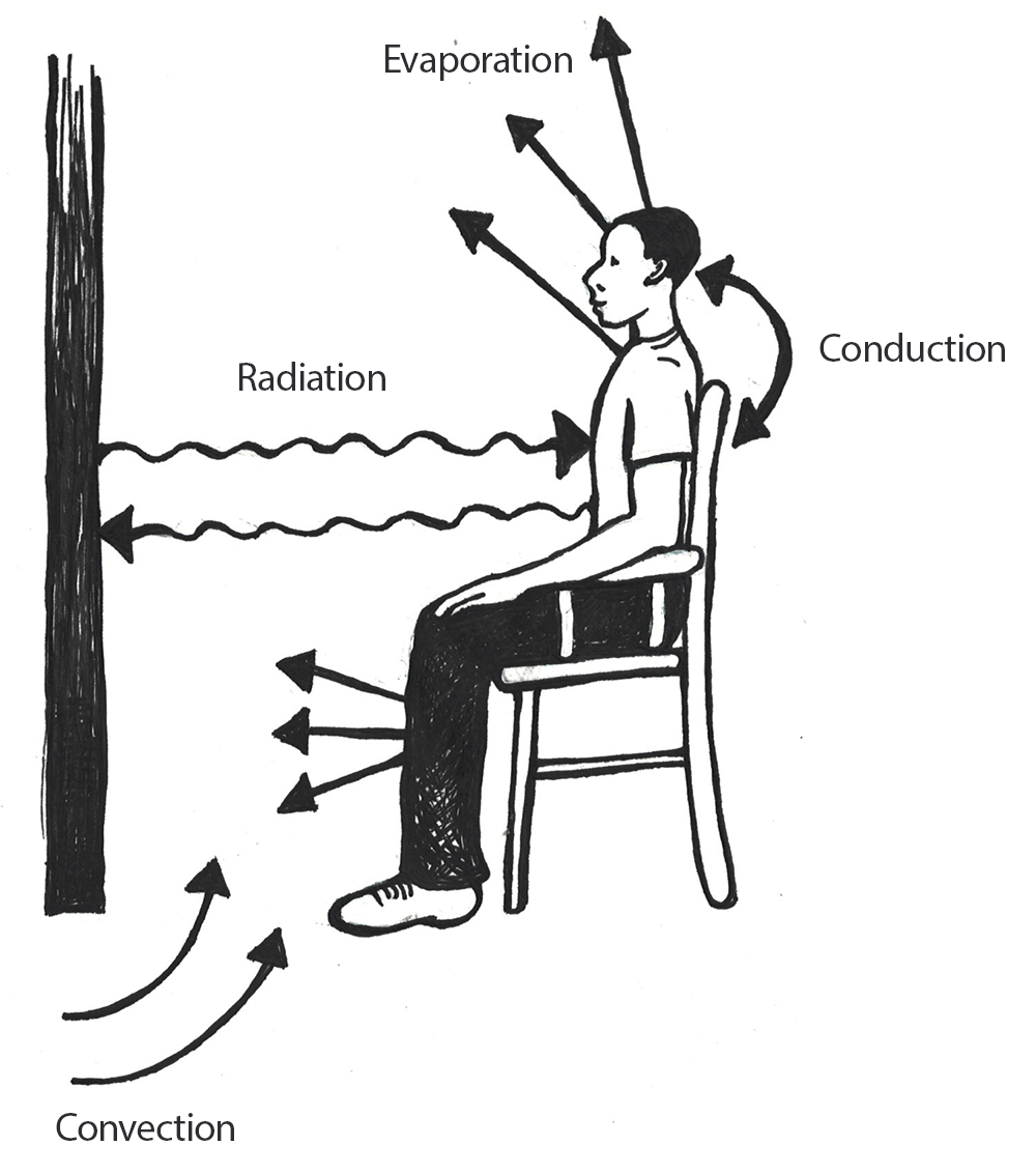
Through conduction processes, heat will move from a warmer body to a cooler one through direct contact. An example of this is when you accidentally touch a hot cooktop with your hand and the heat is transferred from the cooktop to your skin.
With convection, when a warm body is surrounded by a cooler fluid (e.g., air or water), heat will be transferred from the warmer body to the cooler fluid. This is why we will often employ the behavioral adjustment of wearing multiple layers of clothing during the winter in an effort to prevent heat loss to the cooler atmosphere. Conversely, if your body temperature is cooler than that of the air surrounding you, your body will absorb heat.
Depending on your physical condition, most people will begin to sweat around 37.2℃ to 37.7℃ (98.9℉–99.9℉). Sweating is an example of evaporation, which occurs when a liquid, such as the water within our bodies, is converted to a gas. Phase conversions, such as those underlying the evaporative processes of transforming liquids to gasses, require energy. In evaporation, this energy is in the form of heat, and the effect is to cool the body.
The final mechanism for heat loss within the human body is radiation, through which energy in the form of electromagnetic waves is produced at a wavelength that typically lies below that which is visible to the human eye. Although humans gain and lose heat from their bodies through radiation, this form of heat transfer is not visible. Humans are capable of losing and gaining heat through conduction, convection, and radiation; however, heat may not be gained through evaporation.
As the ambient temperature decreases, it becomes increasingly difficult for the human body to regulate its core temperature, which is central to the maintenance of homeostasis. When an individual’s body temperature falls below 34.4℃ (93.9°F), the brain’s hypothalamus becomes impaired, leading to issues with body temperature control. A total loss of the ability to regulate body temperature occurs around 29.4℃ (84.9°F), which may result in death. When the ambient temperature falls below the critical temperature of 31℃ (87.8°F), a nude human body that is at rest will respond with a series of physiological changes to preserve homeostasis (Figure 14.3).
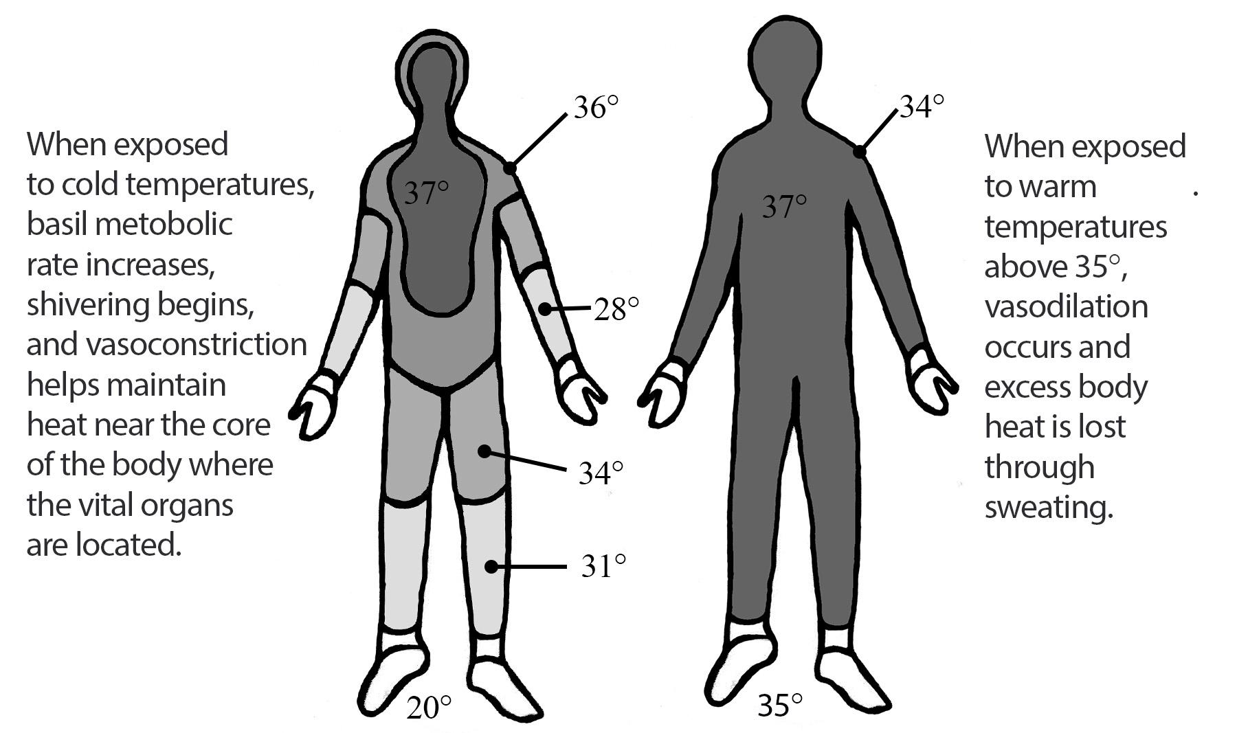
The human body experiences two main types of physiological responses to colder temperatures: those that increase the production of heat and those that seek to retain heat. The production of heat within the body is accomplished through short-term increases in the body’s basal metabolic rate, such as shivering to increase muscular metabolism. An organism’s basic metabolic rate is a measure of the energy required to maintain necessary body processes when the organism is at rest. Increases in basal metabolic rates, such as when we shiver from the cold, require increased consumption of energy-providing nutrients. Of course, such increases in metabolic rates are not infinite, as we may only consume a finite amount of nutrients. As with all acclimatory adjustments, an increase in the basal metabolic rate is merely temporary.
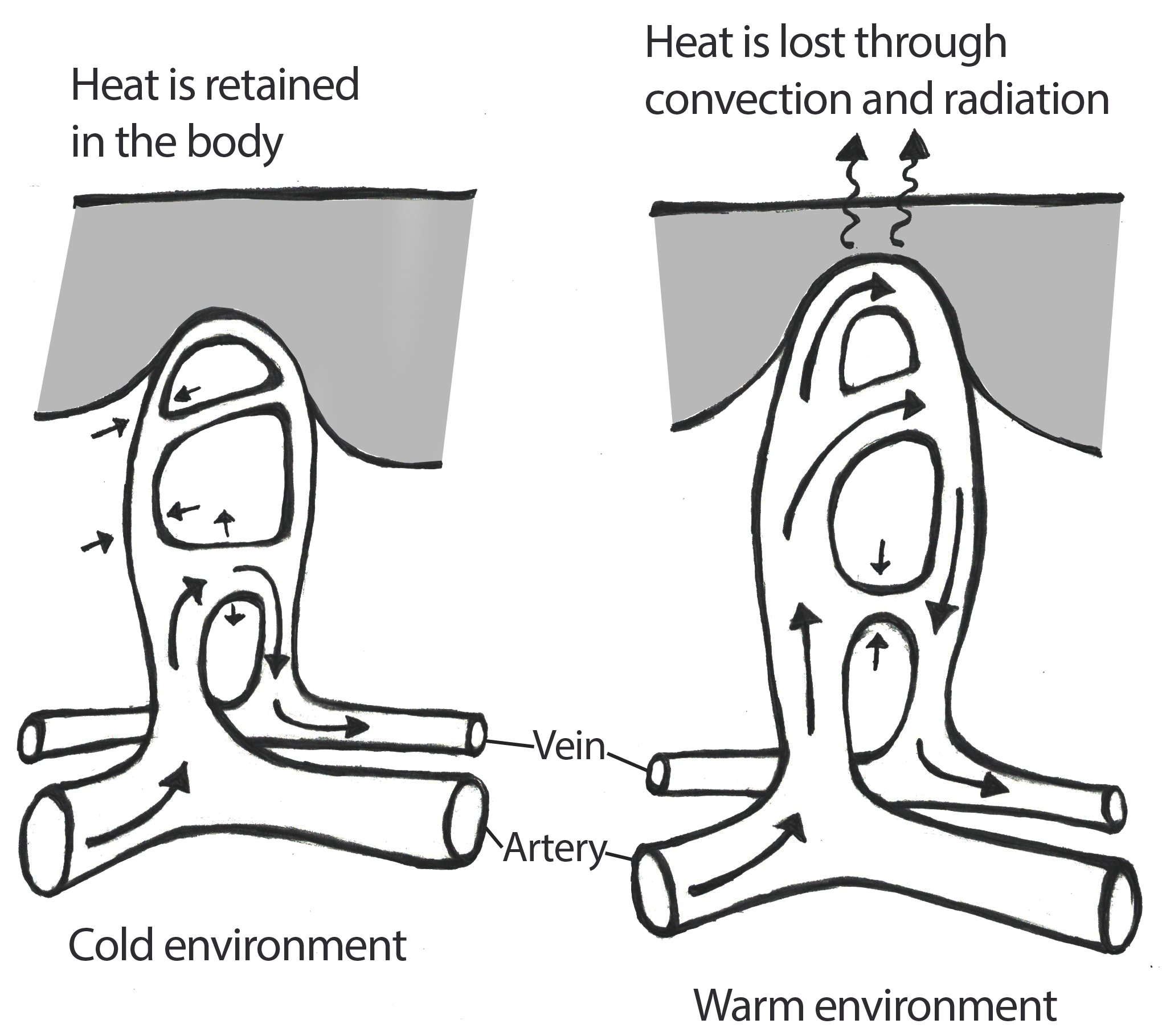
Of the physiological mechanisms to preserve heat already in the body, the most notable is vasoconstriction, or the constriction of peripheral capillaries in the skin. The decreased surface area of the capillaries through vasoconstriction results in less heat reaching the surface of the skin where it would be dissipated into the atmosphere. Vasoconstriction also leads to the maintenance of heat near the core of the body where the vital organs are located. As a trade-off, though, individuals are more at risk of cold-related injuries, such as frost-bite, which can lead to tissue necrosis (tissue death) in regions of the body that are most distant from the core (e.g., fingers, toes, nose, ears, cheeks, chin, etc.).
Just as cold stress presents challenges to maintaining homeostasis, heat does as well. In hot climates, the body will absorb heat from its surroundings (through conduction, convection, and radiation), resulting in potential heat-related disorders, such as heat exhaustion. When the human body is exposed to ambient temperatures above 35℃ (95°F), excess body heat will be lost primarily through evaporative processes, specifically through sweating. All humans, regardless of their environment, have approximately the same number of sweat glands within their bodies. Over time, individuals living in hot, arid environments will develop more sensitive forms of sweat glands resulting in the production of greater quantities of sweat (Best, Lieberman, and Kamilar 2019; Pontzer et al. 2021). In an effort to prevent dehydration due to this form of acclimatory adjustment, there will be an additional reduction in the volume of urine produced by the individual (Pontzer et al. 2021).
As noted in the previous section, some cultural groups, particularly those in equatorial regions, add pungent spices to their foods to inhibit the colonization of bacteria (Sherman and Billing 1999). Although adding spices to decrease spoilage rates is a behavioral adjustment, the application of some forms of peppers triggers an acclimatory adjustment as well. Compounds referred to as capsaicinoids are the secondary byproducts of chili pepper plants’ metabolism and are produced to deter their consumption by some forms of fungi and mammals. When mammals, such as humans, consume the capsaicinoids from chili peppers, a burning sensation may occur within their mouths and along their digestive tracts. This burning sensation is the result of the activation of capsaicin receptors along the body’s nerve pathways. Although the peppers themselves may be at ambient temperature so their consumption is not causing any form of body temperature increase, the human body perceives the pepper as elevating its core temperature due to the activation of the capsaicin receptors. This causes the hypothalamus to react, initiating sweating in an attempt to lower body temperature and maintain homeostasis. The increased piquancy (application of pungent spices to food) as a means of inhibiting food-borne bacterial colonization in warm climates, as well as spices’ ability to trigger sweating processes as a method for cooling the body, is an example of the intersection between behavioral and acclimatory adjustments that utilized within certain populations.
In addition to increased sweat production to maintain homeostasis in excess heat, vasodilation may occur (Figure 14.4). Vasodilation is an expansion of the capillaries within the skin leading to a more effective transfer of heat from within the body to the exterior to allow conductive, convective, radiative, and evaporative (sweating) processes to occur.
Physiologically based acclimatory adjustments to hot, dry climates may be complemented by behavioral adjustments as well. For example, individuals in such climates may limit their physical activity during the times of day when the temperature is typically the hottest. Additionally, these individuals may wear loose-fitting clothing that covers much of their skin. The looseness of the clothing allows for air to flow between the clothing and the skin to permit the effective evaporation of sweat. Although it may seem counterintuitive to cover one’s body completely in a hot climate, the covering of the skin keeps the sun’s rays from directly penetrating the skin and elevating the body’s core temperature.
Acclimatory Adjustments: Altitudinal Stressors
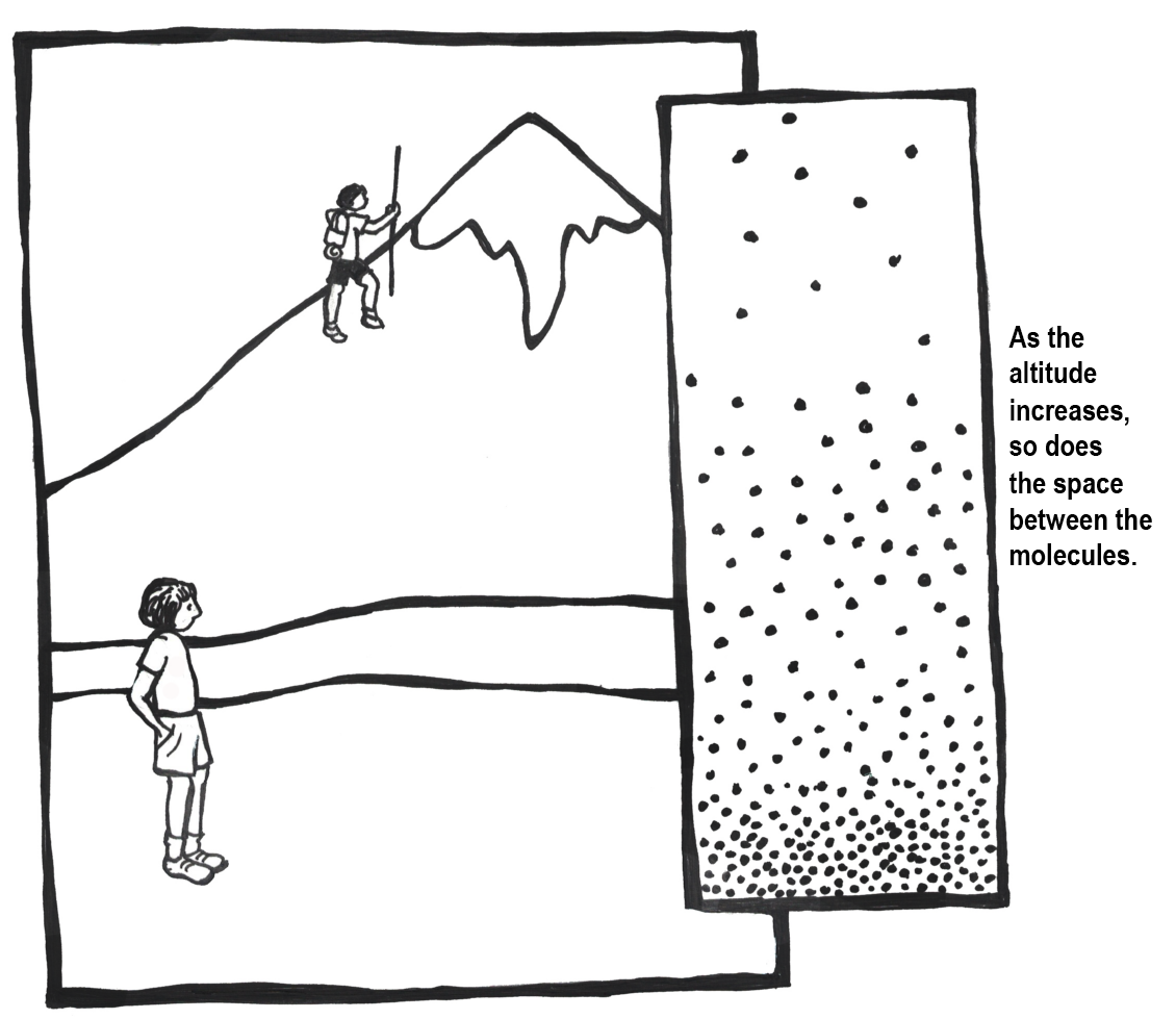
The challenges posed by thermal conditions are but one form of environmental stressor humans must face. High-altitude environments, which are defined as altitudes in excess of 2,400 meters above sea level (masl) or 7,874 feet above sea level (fasl), pose additional challenges to the maintenance of homeostasis in humans. Some of the main stressors encountered by those living within high-altitude environments include decreased oxygen availability, cold temperatures, low humidity, high wind speed, a reduced nutritional base, and increased solar radiation levels. Of these challenges, the most significant is the decreased availability of oxygen.
To visualize how altitude affects the availability of oxygen, imagine two balloons that are each filled with the same quantity of oxygen molecules. One of these balloons is positioned at sea-level and the other is placed high upon a mountain peak. For the balloon at sea level, there is more atmospheric pressure pressing down on the molecules within this balloon. This leads to the oxygen molecules within the sea level balloon being forced into a more compact organization. In contrast, the mountain peak balloon has less atmospheric pressure pressing down on it. This leads to the oxygen molecules within that balloon spreading out from each other since they are not being forced together quite as strongly. This example highlights the availability of oxygen molecules in each breath than we take in low- versus high-altitude environments. At 5,500 masl (approximately 18,000 fasl), the atmospheric pressure is approximately 50% of its value at sea level (Peacock 1998). At the peak of Mount Everest (8,900 masl or approximately 29,200 fasl), the atmospheric pressure is equivalent to only about 30% of their sea level amounts (Peacock 1998; Figure 14.5).
Due to decreased availability of oxygen at higher altitudes, certain acclimatory adjustments are required to ensure the maintenance of homeostasis for individuals other than those who were gestated, born, and raised at high altitude. For these people, their rate of breathing will increase to permit greater quantities of air containing oxygen into the lungs when they ascend into higher altitude environments. An increased speed and depth of breathing, which is referred to as hyperpnea, is not sustainable indefinitely; thus, the rate of breathing begins to decrease as the person becomes acclimatized to the altitude. During the initial phases of high-altitude-related hyperpnea, the heart begins to beat faster but the amount of blood pushed through during each beat decreases slightly. In addition, the body will divert energy from noncritical bodily functions, such as digestive processes.
Once the atmospheric oxygen reaches the alveoli (small air sacs) in the lungs, it spreads across the alveolar membrane and enters erythrocytes (red blood cells). As oxygen reaches the alevoli’s erythrocytes, it loosely binds with hemoglobin (an iron-rich protein) contained in the erythrocytes. When the erythrocytes carrying the hemoglobin-bound oxygen molecules reach capillaries where the partial pressure of oxygen is relatively low, oxygen will be released by the hemoglobin so that it is free for diffusion into body cells. Similar to acclimatory adjustments related to thermal conditions (e.g., shivering or sweating), those related to high altitude may not be infinitely sustained due to their energetically expensive nature.
Although the long-term acclimatory adjustments that an individual from low altitude experiences in a high-altitude environment may permit them to reside there successfully, reproduction within such settings is frequently complicated. With increased altitude comes an increased risk of miscarriage, lower birth weights, and higher infant mortality rates. As the pregnant person’s body seeks to preserve its own homeostasis, there is often a decreased rate and volume of blood flow to the uterus as compared to a pregnant person of similar physiological condition at a lower altitude (Moore, Niermeyer, and Zamudio 1998). This results in a decrease in the amount of oxygen that will be passed through the uterus and placenta to the developing fetus. In addition, pregnant people who experience pregnancy at higher altitudes are more prone to developing preeclampsia (severe elevation of blood pressure), which is linked to increased rates of both fetal and maternal death (Moore, Niermeyer, and Zamudio 1998; Figure 14.6).
Developmental Adjustments
Developmental adjustments occur only in individuals who spent their developmental period (i.e., childhood and adolescence) within a high-altitude environment; they do not apply to those who moved into these environments in the post developmental (i.e., adult) phase. Furthermore, the degree of developmental adjustment within an individual is directly related to their underlying phenotypic plasticity as well as the amount of time during the crucial growth and development period that the individual resides within the challenging environment. Although humans have the remarkable capacity to develop and survive within environments that are not overly conducive to the successful maintenance of homeostasis, there are definitely physiological costs associated with this ability.
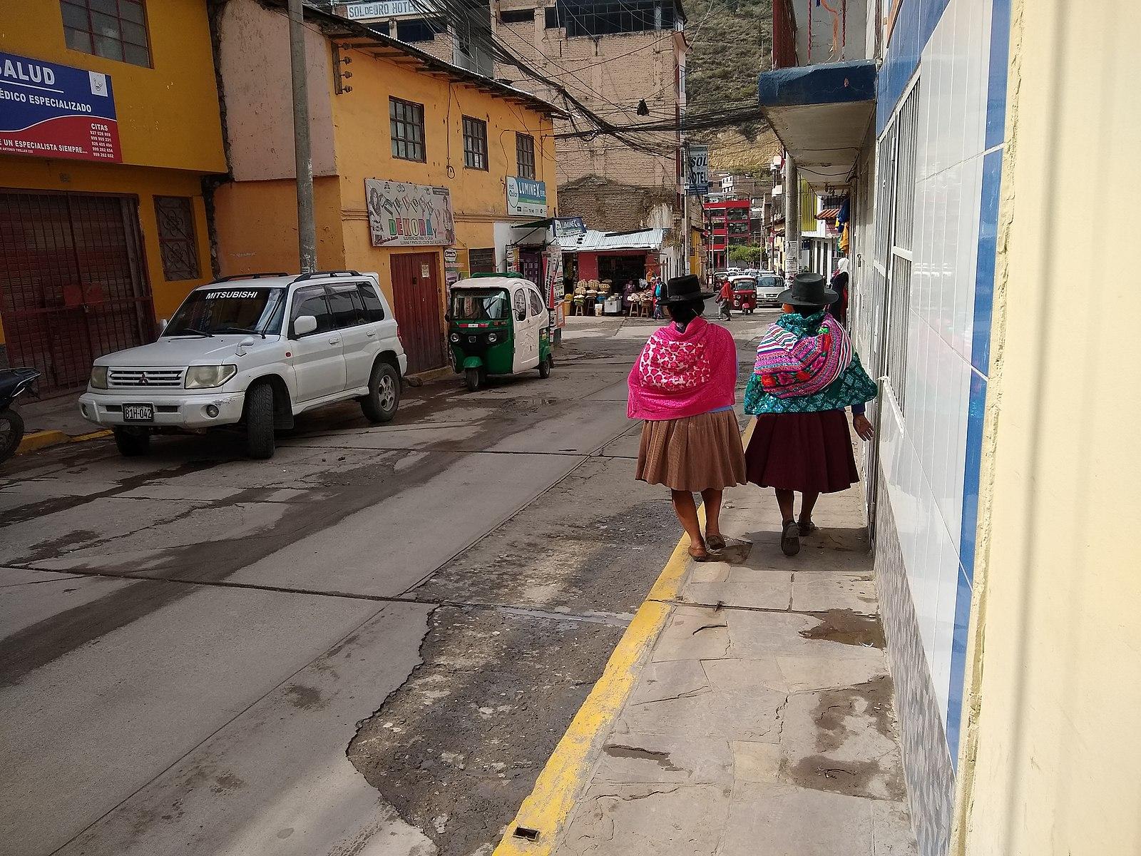
In general, high-altitude natives tend to grow more slowly and physically mature later than their low-altitude counterparts (Figure 14.7). Lowered growth and maturity rates are linked not only to the increased physiological demands placed on the body due to the decreased partial pressure of oxygen but also to reductions in the quality of the nutritional base at higher altitudes. Increased terrain complexity, elevated solar radiation levels, and higher wind speeds coupled with the lower temperatures and humidity levels found at high altitudes leads to difficulties with growing and maintaining crops and raising livestock. Overall, as altitude rises, the quality of the available nutritional base goes down, which is correlated to a lack of the nutrients necessary to ensure proper physiological growth and development in humans. Thus, even though individuals may be able to develop and grow within high-altitude environments, they may not reach their full genetically mediated growth potential as they would in a lower-altitude environment.
Not all developmental adjustments are linked to environmental pressures such as climate or altitude; rather, some of these adjustments are correlated to sociocultural or behavioral practices. Some of these adjustments may affect the physiological appearance of an individual when they are practiced consistently during the development and growth phases.
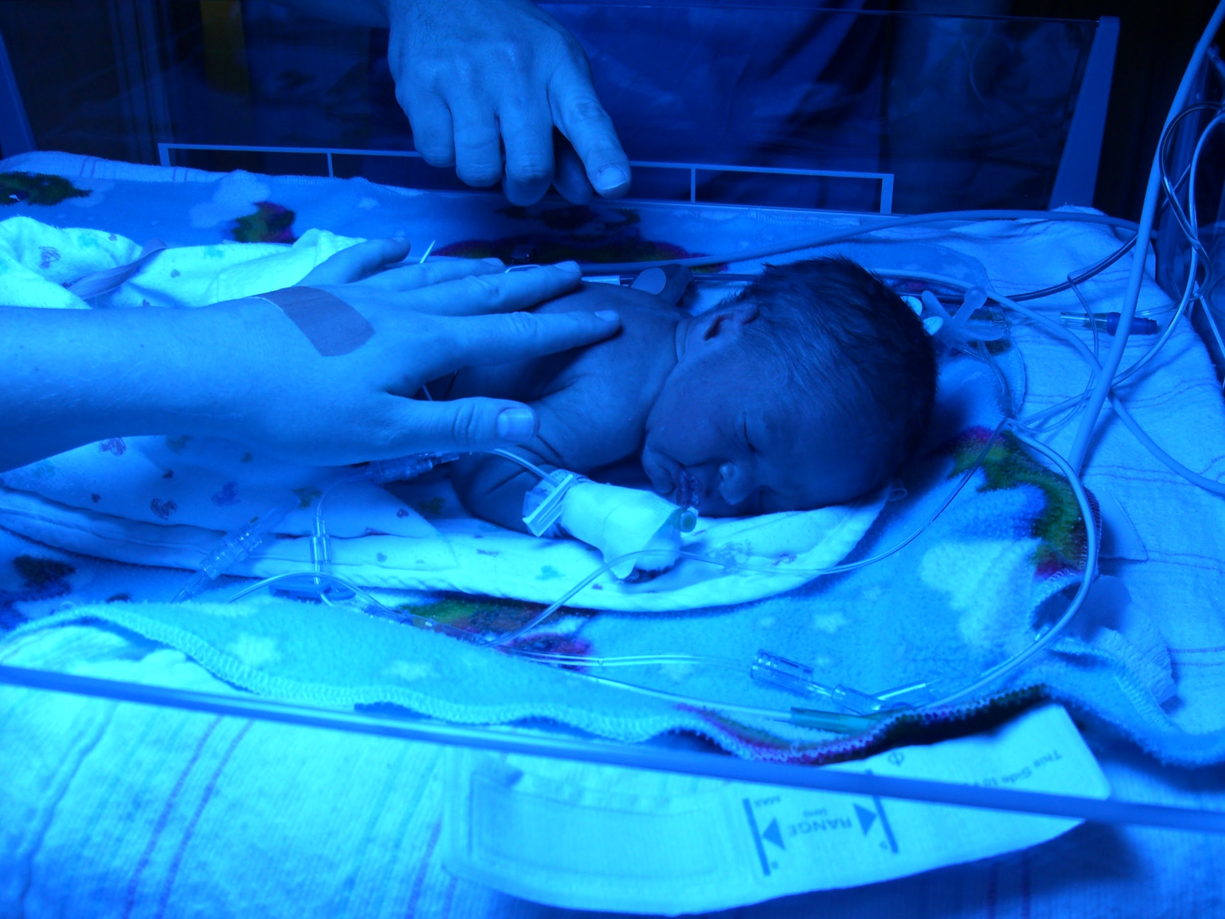
Sudden infant death syndrome (SIDS) has no definitive cause; however, the American Academy of Pediatrics published a report in 1992 linking SIDS to infants (under the age of one) sleeping on their stomachs. The “Back to Sleep” campaign championed by the American Academy of Pediatrics helped educate members of the medical community as well as the public that the best sleep position for infants is on their backs (American Academy of Pediatrics 2000).
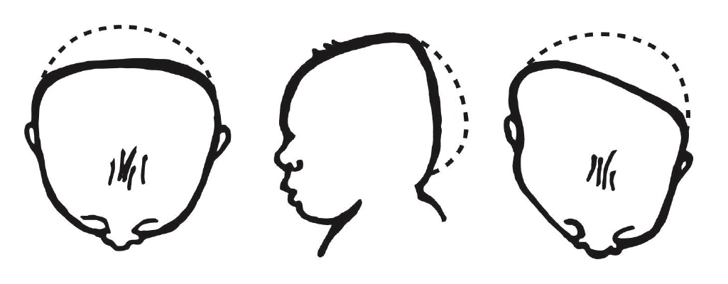
Placing infants on their backs to sleep has led to decreased infant mortality (death) rates due to SIDS; however, it has led to an unintended consequence: infant cranial deformation. The cranial deformations experienced by infants who sleep solely on their back tend to manifest in one of two forms: brachycephaly and plagiocephaly (Roby et al. 2012; Figure 14.8). With positional brachycephaly, the back of the infant’s head appears rather uniformly flattened due to repetitive contact with a flat surface, such as a crib mattress or car seat back. In cases of positional plagiocephaly, the back of the infant’s head appears asymmetrically flattened. This asymmetry is typically due to an uneven distribution of mechanical forces resulting from the manner in which the infant’s head is in contact with a flat surface. The forms of cranial deformation resulting from sleep positioning do not affect the infant’s brain development. For many individuals, the appearance of the deformation is minimized during later development. Still, some individuals will maintain the pattern of cranial deformation acquired during their infancy throughout their lives. The unintentional cranial deformation resulting from placing infants on their backs to sleep as a means of preventing SIDS-related deaths is a physiological indicator of a behavioral adjustment.
Adaptations
As we have just explored, survival and reproduction at high altitudes present numerous physiological challenges for most humans. The behavioral, acclimatory, and developmental adjustments discussed above are all related to the phenotypic plasticity of the individual; however, most adjustments are temporary in nature and they affect a single individual rather than all individuals within a population. But what if the physiological changes were permanent? What if they affected all members of a population rather than just a single individual? The long-term, microevolutionary (i.e., genetic) changes that occur within a population in response to an environmental stressor are referred to as an adaptation. From an evolutionary standpoint, the term adaptation refers to a phenotypic trait (i.e., physiological/morphological feature or behavior) that has been acted upon by natural selection processes to increase a species’ ability to survive and reproduce within a specific environment. Within the field of physiology, the term adaptation refers to traits that serve to restore homeostasis. The physiology-based interpretation of adaptations presumes that all traits serve a purpose and that all adaptations are beneficial in nature; however, this may be a fallacy, since some traits may be present without clear evidence as to their purpose. As such, during the following discussion of various forms of adaptations in human populations, we will focus our attention on phenotypic traits with an evidence-based purpose.
Adaptation: Altitudinal Adaptation
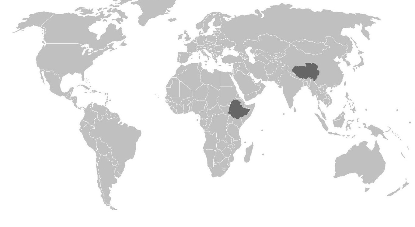
As mentioned in the previous section, there is genomic research supporting the evolutionary selection of certain phenotypes and their corresponding genotypes within indigenous high-altitude populations across the globe. The following discussion focuses on two high-altitude indigenous populations from Tibet and Ethiopia (Figure 14.9). Although these populations share many common genetic traits based on relatively similar evolutionary histories influenced by similar environmental stressors, there is support for local genetically based adaptation as well, based on different genes being acted upon by environmental stressors that may be unique to Tibet and Ethiopia (Bigham 2016).
Tibetan populations have resided in the Tibetan Plateau and Himalayan Mountain regions at elevations exceeding 4,000 masl (13,100 fasl) for at least the past 7,400 years (Meyer et al. 2017). There is evidence of a genetic exchange event involving Tibetan populations and Denisovans around 48,700 years ago, which introduced a haplogroup involving mutations of the EPAS1 gene (Zhang et al. 2021). The EPAS1 is involved in the regulation of erythrocytes and hemoglobin. For individuals originating in lower-altitude environments, EPAS1 stimulates increased erythrocyte production in high-altitude environments as a temporary acclimatory adjustment. For indigenous high-altitude populations of Tibet, the EPAS1 gene mutation introduced by Denisovan introgression inhibits increased erythrocyte production, which reduces potential negative effects (e.g., stroke or heart attack) associated with long-term high levels of erythrocyte production (Gray et al. 2022; Zhang et al. 2021). The erythrocyte count of high-altitude Tibetans with the EPAS1 point mutation is about the same as for individuals residing at sea level.
Populations indigenous to the Semien Plateau of Ethiopia, such as the Oromo and Amhara, share a similar but not identical EPAS1 point mutation with the Tibetan population (Bigham 2016); however, there is no indication that this mutation was derived from Denisovan introgression. The EPAS1 mutations occurred independently from each other; however, their effects are still similar in that they permit the Tibetan and Ethiopian populations to survive at high altitudes. Not all adaptations are related to life in high-altitude environments, however. In the following sections, we will address two more general examples of adaptation in human populations: variations in skin color and differences in body build.
Adaptation: Skin Tone Basics
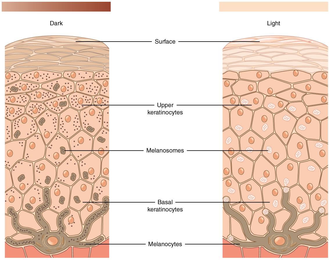
When you think about your own skin tone and compare it to members of your family, do you all possess exactly the same shade? Are some members of your family darker than others? What about your friends? Your classmates? Skin tone occurs along a continuum, which is a reflection of the complex evolutionary history of our species. The expression of skin tone is regulated primarily by melanin and hemoglobin. Melanin is a dark brown-black pigment that is produced by the oxidation of certain amino acids (e.g., tyrosine, cysteine, phenylalanine) in melanocytes. Melanocytes are specialized cells located in the base layer (stratum basale) of the skin’s epidermis as well as several other areas within the body (Figure 14.10). Within the melanocytes, melanin is produced in the special organelle called a melanosome. Melanosomes serve as sites for the synthesis, storage, and transportation of melanin. Melanosomes transport the melanin particles through cellular projections to epidermal skin cells (keratinocytes) as well as to the base of the growing hair root. In the eye, however, melanin particles produced by the melanosomes remain present within the iris and are not transported beyond their origin location. The two main forms of melanin related to skin, hair, and eye color are eumelanin and pheomelanin. All humans contain both eumelanin and pheomelanin within their bodies; however, the relative expression of these two forms of melanin determines an individual’s overall coloring. Eumelanin is a brown-to-black colored melanin particle while pheomelanin is more pink-to-red colored. Individuals with darker skin or hair color have a greater expression of eumelanin than those with lighter-colored skin and blonde or red hair.
Adaptation: Melanogenesis
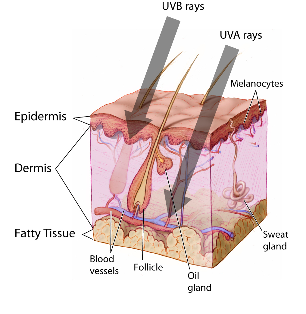
Although all humans have approximately the same number of melanocytes within their epidermis, the production of melanin by these melanocytes varies. There are two forms of melanogenesis (the process through which melanocytes generate melanin): basal and activated. As discussed previously, the expression of eumelanin and pheomelanin by the melanocytes is genetically regulated through the expression of specific receptors (e.g., MC1R) or other melanocyte components (e.g., MFSD12). Basal melanogenesis is dependent upon an individual’s inherent genetic composition and is not influenced by external factors. Activated melanogenesis occurs in response to ultraviolet radiation (UV) exposure, specifically UV-B (short UV wave) exposure. Increased melanogenesis in response to UV-B exposure serves to provide protection to the skin’s innermost layer called the hypodermis, which lies below the epidermis and dermis (Figure 14.11). Melanin in the skin, specifically eumelanin, effectively absorbs UV-B radiation from light—meaning that it will not reach the hypodermal layer. This effect is often more apparent during periods of the year when people tend to be outside more and the weather is warmer, which leads to most donning fewer protective garments. The exposure of skin to sunlight is, of course, culturally mediated with some cultures encouraging the covering of skin at all times.
As previously noted in this chapter, hemoglobin is an iron-rich protein that binds with oxygen in the bloodstream. For individuals with lighter-colored skin, blood vessels near the surface of the skin and the hemoglobin contained within those vessels is more apparent than in individuals with darker skin. The visible presence of hemoglobin coupled with the pink-to-red tone of the pheomelanin leads to lighter-skinned individuals having a pale pink skin tone. Individuals with lighter skin more readily absorb UV radiation as their basal melanin expression is directed more toward the production of pheomelanin than eumelanin. But why are there so many variations in skin tone in humans? To answer this question, we now turn toward an exploration of an evolutionary-based adaptation of skin tone as a function of the environment.
Adaptation: Evolutionary Basis for Skin Tone Variation
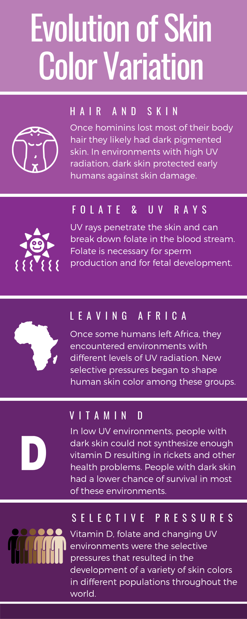
Skin cancer is a significant concern for many individuals with light skin tone as the cumulative exposure of the epidermis and underlying skin tissues to UV radiation may result in the development of abnormal cells within those tissues, leading to malignancies. Although darker-skinned individuals are at risk for skin cancer as well, they are less likely to develop it due to increased levels of melanin, specifically eumelanin, in their skin. Even though skin cancer is a serious health concern for some individuals, most skin cancers occur in the postreproductive years; therefore, it is improbable that evolutionary forces favoring varying melanin expression levels are related to a selective pressure to avoid such cancers. Furthermore, if avoiding skin cancer were the primary factor driving the evolution of various skin tones, then it reasons that everyone would have the most significant expression of eumelanin possible. So, why do we have different skin tones (Figure 14.12)?
The term cline (introduced in Chapter 13) refers to the continuum or spectrum of gradations (i.e., levels or degrees) from one extreme to another. With respect to skin tone, the various tonal shades occur clinally with darker skin being more prevalent near the equator and gradually decreasing in tone (i.e., decreased melanin production) in more distant latitudes. For individuals who are indigenous to equatorial regions, the increased levels of melanin within their skin provides them with a measure of protection against both sunburn and sunstroke because the melanin is more reflective of UV radiation than hemoglobin. In cases of severe sunburn, eccrine glands are affected, resulting in an individual’s ability to sweat being compromised. As sweat is the body’s most effective means of reducing its core temperature to maintain homeostasis, damage to the eccrine glands may lead to numerous physiological issues related to heat that may ultimately result in death.
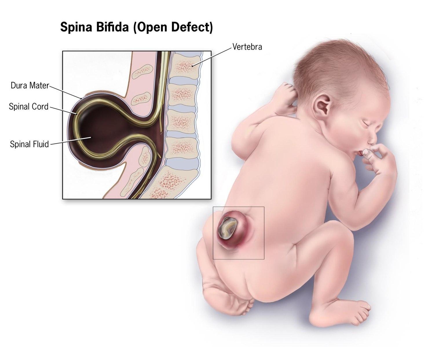
Even though avoiding severe sunburn and sunstroke is of great importance to individuals within equatorial regions, this is likely not the primary factor driving the evolutionary selection of darker skin within these regions. It has been proposed that UV radiation’s destruction of folic acid, which is a form of B-complex vitamin, may have led to the selection of darker skin in equatorial regions. For pregnant people, low levels of folic acid within the body during gestation may lead to defects in the formation of the brain and spinal cord of the fetus. This condition, which is referred to as spina bifida (Figure 14.13), often significantly reduces an infant’s chances of survival without medical intervention. In people producing sperm, low levels of folic acid within the body reduce sperm quantity and quality. Thus, in geographic regions with high UV radiation levels (i.e., equatorial regions), there appears to be an evolutionarily driven correlation between darker skin and fertility.
If darker skin tone is potentially correlated to more successful reproduction, then why do lighter shades of skin exist? One hypothesis is that there is a relationship between lighter skin tone and vitamin D synthesis within the body. When skin is exposed to the UV-B radiation waves in sunlight, a series of chemical reactions occur within the epidermis leading to the production of vitamin D3, which is a fat-soluble vitamin that assists the body with absorbing calcium and phosphorus in the small intestine. These nutrients are among those that are critical for the proper growth and maintenance of bone tissue within the body. In the absence of adequate minerals, particularly calcium, bone structure and strength will be compromised, leading to the development of rickets during the growth phase. Rickets is a disease affecting children during their growth phase. It is characterized by inadequately calcified bones that are softer and more flexible than normal. Individuals with rickets will develop a true bowing of their legs, which may affect their mobility (Figure 14.14). In addition, deformation of pelvic bones in people who may become pregnant may occur as a result of rickets, leading to complications with reproduction. In adults, a deficiency in vitamin D3 will often result in osteomalacia, which is a general softening of the bones due to inadequate mineralization. As noted, a variety of maladies may occur due to the inadequate production or absorption of vitamin D3, as well as the destruction of folate within the human body. Therefore, from an evolutionary perspective, natural selection should favor a skin tone that is best suited to a given environment.

In general, the trend related to lighter skin pigmentation further from the equator follows a principle called Gloger’s Rule. This rule states that within the same species of mammals the more heavily pigmented individuals tend to originate near the equator while lighter-pigmented members of the species will be found in regions further from the equator. Gloger’s Rule applies latitudinally; however, it does not appear to hold for certain human populations near the poles. Specifically, it does not apply to the Inuit people (Figure 14.15), who are indigenous to regions near the North Pole and currently reside in portions of Canada, Greenland, Alaska, and Denmark. The Inuit have a darker skin tone that would not be anticipated under the provisions of Gloger’s Rule. The high reflectivity of light off of snow and ice, which is common in polar regions, necessitates the darker skin tone of these individuals to prevent folic acid degradation just as it does for individuals within equatorial regions. The consumption of vitamin D–rich foods, such as raw fish, permits the Inuit to reside at high latitudes with darker skin tone while preventing rickets.
Adaptation: Shape and Size Variations
In addition to natural selection playing a role in the determination of melanin expression, it plays a significant role in the determination of the shape and size of the human body. As previously discussed, the most significant thermodynamic mechanism of heat loss from the body is radiation. At temperatures below 20℃ (68℉), the human body loses around 65% of its heat to radiative processes; however, the efficiency of radiation is correlated to the overall body shape and size of the individual. There is a direct correlation between the ratio of an object’s surface area to mass and the amount of heat that may be lost through radiation. For example, two objects of identical composition and mass are heated to the same temperature. One object is a cube and the other is a sphere. Which object will cool the fastest? Geometrically, a sphere has the smallest surface area per unit mass of any three-dimensional object, so the sphere will cool more slowly than the cube. In other words, the smaller the ratio of the surface area to mass an object has, the more it will retain heat. With respect to the cube in our example, mass increases by the cube, but surface area may increase only by the square, so size will affect the mass to surface area ratio. This, in general, holds true for humans, as well.
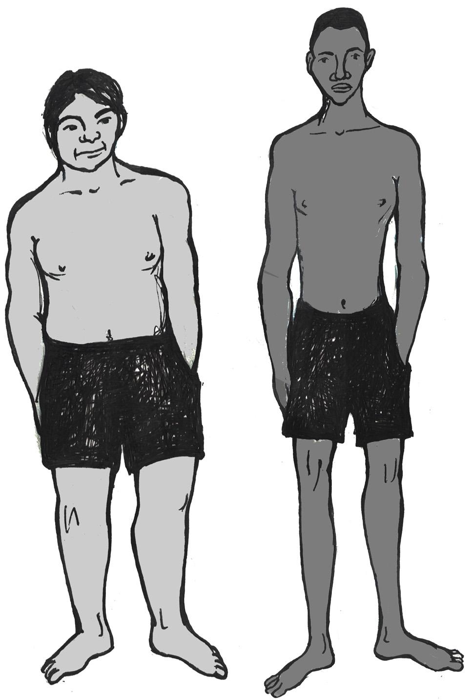
In regions where temperatures are consistently cold, the body shape and size of individuals indigenous to the area tend to be more compact. These individuals have a relatively higher body mass to surface area (i.e., skin) than their counterparts from equatorial regions where the average temperatures are considerably warmer. Individuals from hot climates, such as the Fulani (Figure 14.16a) of West Africa, have limbs that are considerably longer than those of individuals from cold climates, such as the Inuit of Greenland (Figure 14.16b). Evolutionarily, the longer limbs of individuals from equatorial regions (e.g., the Fulani) provide a greater surface area (i.e., lower body mass to surface area ratio) for the dissipation of heat through radiative processes. In contrast, the relatively short limbs of Arctic-dwelling people, such as the Inuit, allows for the retention of heat because there is a decreased surface area through which heat may radiate away from the body.
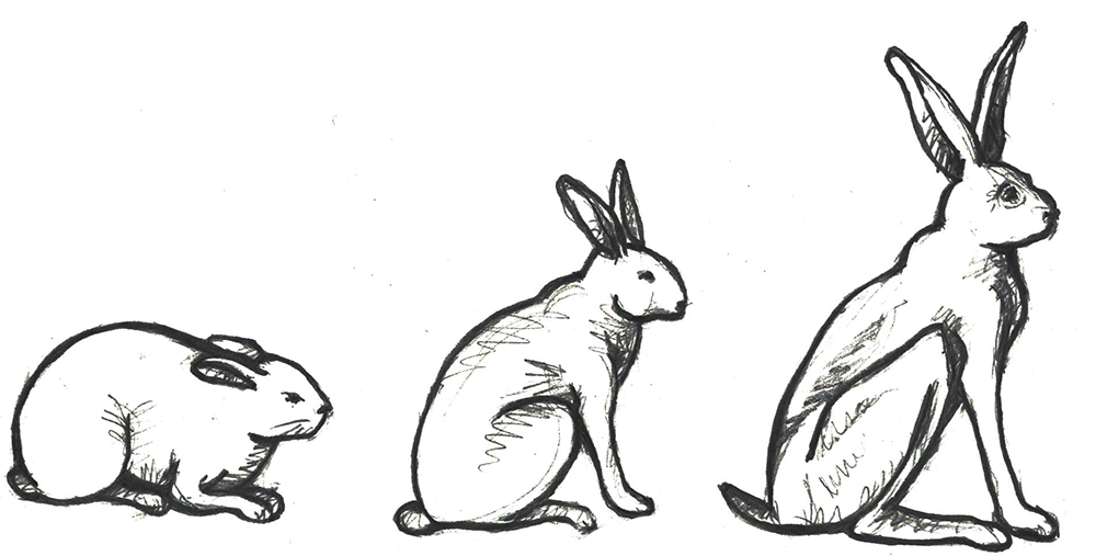
As described above, there are certain trends related to the general shape and size of human bodies in relation to the thermal conditions. To better describe these trends, we turn to a couple of general principles that are applicable to a variety of species beyond humans. Bergmann’s Rule predicts that as average environmental temperature decreases, populations are expected to exhibit an increase in weight and a decrease in surface area (Figure 14.17a). Also, within the same species of homeothermic animals, the relative length of projecting body parts (e.g., nose, ears, and limbs) increases in relation to the average environmental temperature (Figure 14.17b). This principle, referred to as Allen’s Rule, notes that longer, thinner limbs are advantageous for the radiation of excess heat in hot environments and shorter, stockier limbs assist with the preservation of body heat in cold climates. A measure of the crural index (crural index = tibia length ÷ femur length) of individuals from various human populations provides support for Allen’s Rule since this value is lower in individuals from colder climates than it is for those from hot climates. The crural indices for human populations vary directly with temperature, so individuals with higher crural index values are generally from regions with a warmer average environmental temperature. Conversely, the crural indices are lower for individuals from regions where there are colder average temperatures.

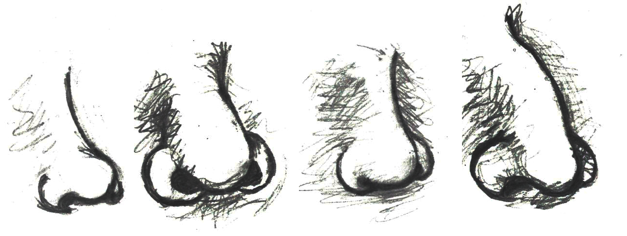
Nasal shape and size (Figure 14.18) is another physiological feature affected by our ancestors’ environments. The selective role of climate in determining human nasal variation is typically approached by dividing climates into four adaptive zones: hot-dry, hot-wet, cold-dry, and cold-wet (Maddux et al. 2016). A principal role of the nasal cavity is to condition (i.e., warm and humidify) ambient air prior to its reaching the lungs. Given this function of the nasal cavity, it is anticipated that different nasal shapes and sizes will be related to varying environments. In cold-dry climates, an individual’s nasal cavity must provide humidification and warmth to the dry air when breathing in through the nose (Noback et al. 2011). Also, in that type of climate, the nasal cavity must conserve moisture and minimize heat loss during when the individual exhales through the nose (Noback et al. 2011). From a physiological stress perspective, this is a stressful event.
Conversely, in hot-wet environments, there is no need for the nasal cavity to provide additional moisture to the inhaled air nor is there a need to warm the air or to preserve heat within the nasal cavity (Noback et al. 2011). So, in hot-wet climates, the body is under less physiological stress related to the inhalation of ambient air than in cold-dry climates. As with most human morphological elements, the shape and size of the nasal cavity occurs along a cline. Due to the environmental stressors of cold-dry environments requiring the humidification and warming of air through the nasal cavity, individuals indigenous to such environments tend to have taller (longer) noses with a reduced nasal entrance (nostril opening) size (Noback et al. 2011). This general shape is referred to as leptorrhine, and it allows for a larger surface area within the nasal cavity itself for the air to be warmed and humidified prior to entering the lungs (Maddux et al. 2016). In addition, the relatively small nasal entrance of leptorrhine noses serves as a means of conserving moisture and heat (Noback et al. 2011). Individuals indigenous to hot-wet climates tend to have platyrrhine nasal shapes, which are shorter with broader nasal entrances (Maddux et al. 2016). Since individuals in hot-wet climates do not need to humidify and warm the air entering the nose, their nasal tract is shorter and the nasal entrance wider to permit the effective cooling of the nasal cavity during respiratory processes.
Adaptation: Infectious Disease
Throughout our evolutionary journey, humans have been exposed to numerous infectious diseases. In the following section, we will explore some of the evolutionary-based adaptations that have occurred in certain populations in response to the stressors presented by select infectious diseases. One of the primary examples of natural selection processes acting on the human genome in response to the presence of an infectious disease is the case of the relationship between the sickle-cell anemia trait and malaria, introduced in Chapter 4.
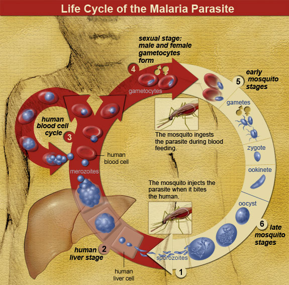
Malaria is a zoonotic disease (an infectious disease transmitted between animals and humans; it is covered in more detail in Chapter 16). It is caused by the spread of the parasitic protozoa from the genus Plasmodium (Figure 14.19). These unicellular, eukaryotic protozoa are transmitted through the bite of a female Anopheles mosquito. During the bite process, the protozoan parasites present within an infected mosquito’s saliva enter a host’s bloodstream where they are transported to the liver. Within the liver, the parasites multiply and are eventually released into the bloodstream, where they infect erythrocytes. Once inside the erythrocytes, the parasites reproduce until they exceed the cell’s storage capacity, causing it to burst and release the parasites into the bloodstream once again. This replication cycle continues as long as there are viable erythrocytes within the host to infect.
General complications from malaria infections include the following: enlargement of the spleen (due to destruction of infected erythrocytes); lower number of thrombocytes (also called platelets, required for coagulation/clotting of blood); high levels of bilirubin (a byproduct of hemoglobin breakdown in the liver) in the blood; jaundice (yellowing of the skin and eyes due to increased blood bilirubin levels); fever; vomiting; retinal (eye) damage; and convulsions (seizures). In 2020, there were 241 million cases of malaria reported globally, with 95% of those cases originating in Africa (World Health Organization 2021). In sub-Saharan Africa, where incidents of malaria are the highest in the world, 125 million pregnancies are affected by malaria, resulting in 200,000 infant deaths (Hartman, Rogerson, and Fischer 2013). Pregnant people who become infected during the gestational process are more likely to have low-birthweight infants due to prematurity or growth restriction inside the uterus (Hartman, Rogerson, and Fischer 2013). After birth, infants born to malaria-infected pregnant people are more likely to develop infantile anemia (low red-blood cell counts), a malaria infection that is not related to the maternal malarial infection, and they are more likely to die than infants born to non-malaria-infected pregnant people (Hartman, Rogerson, and Fischer 2013).
For children and adolescents whose brains are still developing, there is a risk of cognitive (intellectual) impairment associated with some forms of malaria infections (Fernando, Rodrigo, and Rajapakse 2010). Given the relatively high rates of morbidity (disease) and mortality (number of deaths) associated with malaria, it is plausible that this disease may have served as a selective pressure during human evolution. Support for natural selection related to malaria resistance is related to genetic mutations associated with sickle cell, thalassemia, glucose-6-phosphate dehydrogenase (G6PD) deficiency, and the absence of certain antigens (molecules capable of inducing an immune response from the host) on erythrocytes. For the purposes of this text, we will focus our discussion on the relationship between sickle cell disease and malaria.
Sickle cell disease is a group of genetically inherited blood disorders characterized by an abnormality in the shape of the hemoglobin within erythrocytes. It is important to note that there are multiple variants of hemoglobin, including, but not limited to the following: A, D, C, E, F, H, S, Barts, Portland, Hope, Pisa, and Hopkins. Each of these variants of hemoglobin may result in various conditions within the body; however, for the following explanation we will focus solely on variants A and S.
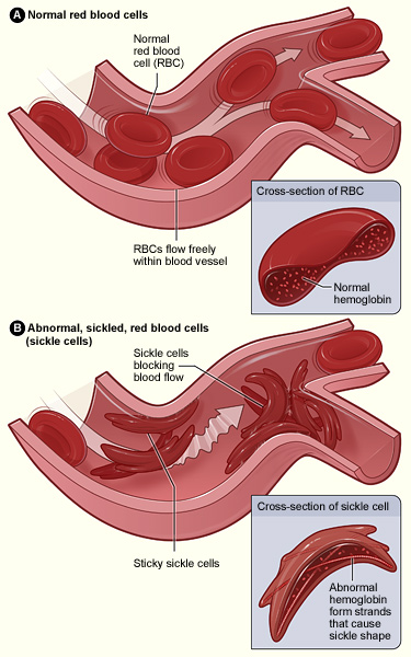
Individuals who inherit a mutated gene (hemoglobin with a sickled erythrocyte variety, HbS) on chromosome 11 from both parents will develop sickle cell anemia, which is the most severe form of the sickle cell disease family (Figure 14.20). The genotype of an individual with sickle cell anemia is HbSS; whereas, an individual without sickle cell alleles has a genotype of HbAA representing two normal adult hemoglobin type A variants. Manifestations of sickle cell anemia (HbSS) range from mild to severe, with some of the more common symptoms being anemia, blood clots, organ failure, chest pain, fever, and low blood-oxygen levels. In high-income countries with advanced medical care, the median life expectancy of an HbSS individual is around 60 years; however, in low-income countries where advanced medical care is scarce, as many as 90% of children with sickle cell disease perish before the age of five (Longo et al. 2017).
Considering that advanced medical care was not available during much of human evolutionary history, it stands to reason that the majority of individuals with the HbSS genotype died before the age of reproduction. If that is the case though, why do we still have the HbS variant present in modern populations? As covered earlier in this textbook, the genotype of an individual is composed of genes from both biological parents. In the case of an individual with an HbSS genotype, the sickle cell allele (HbS) was inherited from each of the parents. For individuals with the heterozygous genotype of HbSA, they have inherited both a sickle cell allele (HbS) and a normal hemoglobin allele (HbA). Heterozygous (HbSA) individuals who reside in regions where malaria is endemic may have a selective advantage. They will experience a sickling of some, but not all, of their erythrocytes. As discussed in the following paragraph, HbSA heterozygous individuals are less likely to die from malaria infections than their HbAA counterparts. Unlike an individual with the HbSS genotype, someone with HbSA may experience some of the symptoms listed above; however, they are generally less severe.
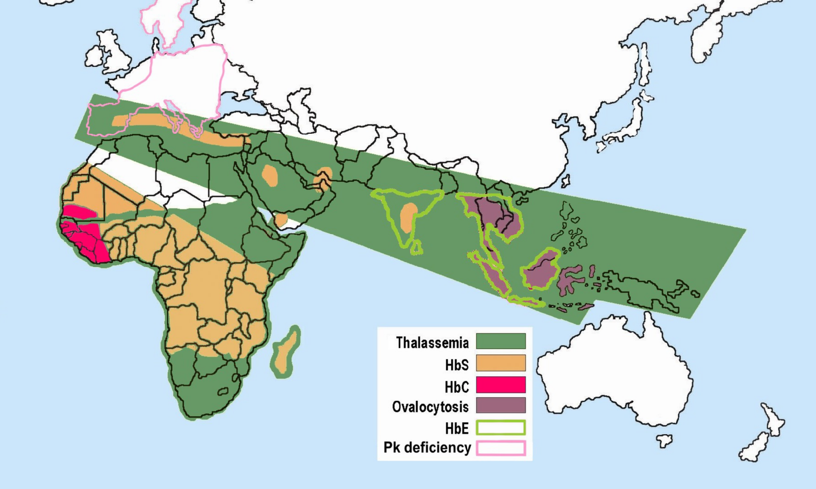
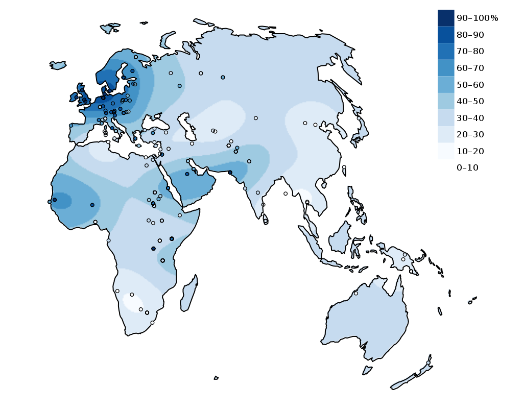
As noted earlier, the mechanism through which Plasmodium protozoan parasites replicate involves human erythrocyte cells. However, due to their sickled shape, as well as the presence of an abnormally shaped protein within the cell, the parasites are unable to replicate effectively in the erythrocyte cells coded for by the HbS allele (Cyrklaff et al. 2011). An individual who has an HbSA genotype and an active malaria infection will become ill with the disease to a lesser extent than someone with an HbAA genotype, which increases their chances of survival. Although normal erythrocytes (regulated by the HbA allele) allow for parasite replication, they are not able to replicate in HbS erythrocytes of the heterozygote. So, individuals with the HbSA genotype are more likely to survive a malaria infection than an individual who is HbAA. Although individuals with the HbSA genotype may endure some physiological complications related to the sickling of some of their erythrocytes, their morbidity and mortality rates are lower than they are for HbSS members of the population. The majority of individuals who are heterozygous or homozygous for the HbS trait have ancestors who originated in sub-Saharan Africa, India, Saudi Arabia, and regions in South and Central America, the Mediterranean (Turkey, Greece, and Italy), and the Caribbean (Centers for Disease Control and Prevention 2017; Figure 14.21).
With respect to the history of these regions, during the early phases of settlement horticulture was the primary method of crop cultivation. Typically performed on a small scale, horticulture is based on manual labor and relatively simple hand tools rather than the use of draft animals or irrigation technologies. Common in horticulture is swidden, or the cutting and burning of plants in woodland and grassland regions. The swidden is the prepared field that results following a slash-and-burn episode. This practice fundamentally alters the soil chemistry, removes plants that provide shade, and increases the areas where water may pool. This anthropogenically altered landscape provides the perfect breeding ground for the Anopheles mosquito, as it prefers warm, stagnant pools of water (Figure 14.22).
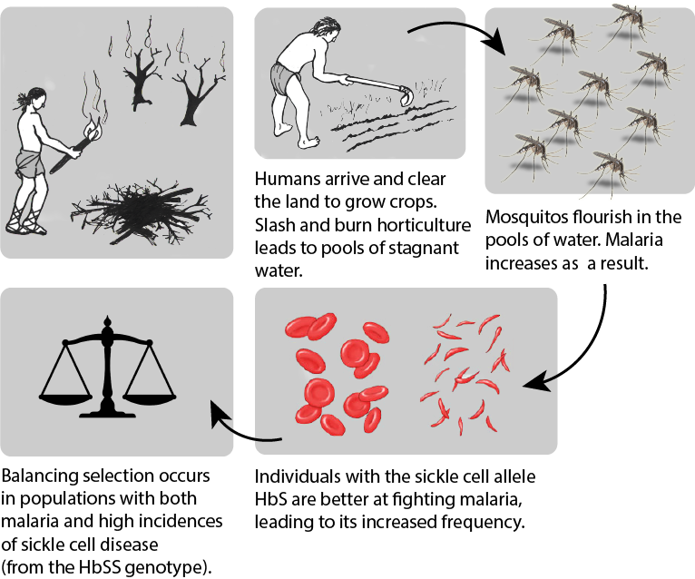
Although swidden agriculture was historically practiced across the globe, it became most problematic in the regions where the Anopheles mosquito is endemic. These areas have the highest incidence rates of malaria infection. Over time, the presence of the Anopheles mosquito and the Plasmodium parasite that it transmitted acted as a selective pressure, particularly in regions where swidden agricultural practices were common, toward the selection of individuals with some modicum of resistance against the infection. In these regions, HbSS and HbSA individuals would have been more likely to survive and reproduce successfully. Although individuals and populations are far more mobile now than they have been throughout much of history, there are still regions where we can see higher rates of malaria infection as well as greater numbers of individuals with the HbS erythrocyte variant. The relationship between malaria and the selective pressure for the HbS variant is one of the most prominent examples of natural selection in the human species within recent evolutionary history.
Adaptation: Lactase Persistence
With the case of sickled erythrocytes and their resistance to infection by malaria parasites, there is strong support for a cause-and-effect-style relationship linked to natural selection. Although somewhat less apparent, there is a correlation between lactase persistence and environmental challenges. Lactase-phlorizin hydrolase (LPH) is an enzyme that is primarily produced in the small intestine and permits the proper digestion of lactose, a disaccharide (composed of two simple sugars: glucose and galactose) found in the milk of mammals. Most humans will experience a decrease in the expression of LPH following weaning, leading to an inability to properly digest lactose. Generally, LPH production decreases between the ages of two and five and is completely absent by the age of nine (Dzialanski et al. 2016). For these individuals, the ingestion of lactose may lead to a wide variety of gastrointestinal ailments, including abdominal bloating, increased gas, and diarrhea. Although the bloating and gas are unpleasant, the diarrhea caused by a failure to properly digest lactose can be life-threatening if severe enough due to the dehydration it can cause. Some humans, however, are able to produce LPH far beyond the weaning period.
Individuals who continue to produce LPH have what is referred to as the lactase persistence trait. The lactase persistence trait is encoded for a gene called LCT, which is located on human chromosome 2 (Ranciaro et al. 2014; see also Chapter 3). From an evolutionary and historical perspective, this trait is most commonly linked to cultures that have practiced cattle domestication (Figure 14.23). For individuals in those cultures, the continued expression of LPH may have provided a selective advantage. During periods of environmental stress, such as a drought, if an individual is capable of successfully digesting cow’s milk, they have a higher chance of survival than someone who suffers from diarrhea-linked dehydration due to a lack of LPH. Although the frequency of the lactase persistence trait is relatively low among African agriculturalists, it is high among pastoralist populations that are traditionally associated with cattle domestication, such as the Tutsi and Fulani, who have frequencies of 90% and 50%, respectively (Tishkoff et al. 2007).
Cattle domestication began around 11,000 years ago in Europe (Beja-Pereira et al. 2006) and 7,500 to 9,000 years ago in the Middle East and North Africa (Tishkoff et al. 2007). Based on human genomic studies, it is estimated that the mutation for the lactase persistence trait occurred around 2,000 to 20,000 years ago for European populations (Tishkoff et al. 2007). For African populations, the lactase persistence trait emerged approximately 1,200 to 23,000 years ago (Gerbault et al. 2011). This begs the question: Is this mutation the same for both populations? It appears that the emergence of the lactase persistence mutation in non-European populations, specifically those in East Africa (e.g., Tutsi and Fulani), is a case of convergent evolution. With convergent evolution events, a similar mutation may occur in species of different lineages through independent evolutionary processes. Based on our current understanding of the genetic mutation pathways for the lactase persistence trait in European and African populations, these mutations are not representative of a shared lineage. In other words, just because a person of European origin and a person of African origin can each digest milk due to the presence of the lactase-persistence trait in their genotypes, it does not mean that these two individuals inherited it due to shared common ancestry.
Is it possible that the convergent evolution of similar lactase-persistence traits in disparate populations is merely a product of genetic drift? Or is there evidence for natural selection? Even though 23,000 years may seem like a long time, it is but a blink of the proverbial evolutionary eye. From the perspective of human evolutionary pathways, mutations related to the LCT gene have occurred relatively recently. Similar genetic changes in multiple populations through genetic drift processes, which are relatively slow and directionless, fail to accumulate as rapidly as lactase-persistence traits (Gerbault et al. 2011). The widespread accumulation of these traits in a relatively short period of time supports the notion that an underlying selective pressure must be driving this form of human evolution. Although to date no definitive factors have been firmly identified, it is thought that environmental pressures are likely to credit for the rapid accumulation of the lactase-persistence trait in multiple human populations through convergent evolutionary pathways.
Special Topic: Skin Tone Genetic Regulation
The melanocortin 1 receptor (MC1R) gene acts to control which types of melanin (eumelanin or pheomelanin) are produced by melanocytes. The MC1R receptor is located on the surface of the melanocyte cells (Quillen et al. 2018). Activation of the MC1R receptors may occur through exposure to specific environmental stimuli or due to underlying genetic processes. Inactive or blocked MC1R receptors result in melanocytes producing pheomelanin. If the MC1R gene receptors are activated, then the melanocytes will produce eumelanin. Thus, individuals with activated MC1R receptors tend to have darker-pigmented skin and hair than individuals with inactive or blocked receptors.
The alleles of another gene, the major facilitator, superfamily domain-containing protein 12 (MFSD12) gene, affect the expression of melanocytes in a different way than the MC1R gene. Instead of affecting the activation of melanocyte receptors, the MFSD12 alleles indirectly affect the membranes of melanocyte lysosomes (Quillen et al. 2018). The melanocyte’s lysosomes are organelles containing digestive enzymes, which ultimately correlate to varying degrees of pigmentation in humans. Variations in the membranes of the melanocyte lysosomes ultimately correlate to differing degrees of pigmentation in humans.
Ancestral MFSD12 allele variants are present in European and East Asian populations and are associated with lighter pigmentation of the skin (Crawford et al. 2017; Quillen et al. 2018). In addition, this ancestral variant is also associated with Tanzanian, San, and Ethiopian populations of Afro-Asiatic ancestry (Crawford et al. 2017; Quillen et al. 2018). In contrast, the more derived (i.e., more recent) allele variants that are linked to darker skin tones are more commonly present in East African populations, particularly those of Nilo-Saharan descent (Crawford et al. 2017; Quillen et al. 2018). The notion that ancestral alleles of MFSD12 are associated with lighter skin pigmentation is in opposition to the commonly accepted idea that our pigmentation was likely darker throughout early human evolution (Crawford et al. 2017; Quillen et al. 2018). Due to the complexity of the human genome, MFSD12 and MC1R are but two examples of alleles affecting human skin tone. Furthermore, there is genetic evidence suggesting that certain genomic variants associated with both darker and lighter skin color have been subject to directional selection processes for as long as 600,000 years, which far exceeds the evolutionary span of Homo sapiens sapiens (Crawford et al. 2017; Quillen et al. 2018).
Human Variation: Our Story Continues
From the time that the first of our species left Africa, we have had to adjust and adapt to numerous environmental challenges. The remarkable ability of human beings to maintain homeostasis through a combination of both nongenetic (adjustments) and genetic (adaptations) means has allowed us to occupy a remarkable variety of environments, from high-altitude mountainous regions to the tropics near the equator. From adding piquant, pungent spices to our foods as a means of inhibiting food-borne illnesses due to bacterial growth to donning garments specially suited to local climates, behavioral adjustments have provided us with a nongenetic means of coping with obstacles to our health and well-being. Acclimatory adjustments, such as sweating when we are warm in an attempt to regulate our body temperature or experiencing increased breathing rates as a means of increasing blood oxygen levels in regions where the partial pressure of oxygen is low, have been instrumental in our survival with respect to thermal and altitudinal environmental challenges. For some individuals, developmental adjustments that were acquired during their development and growth phases (e.g., increased heart and lung capacities for individuals from high-altitude regions) provide them with a form of physiological advantage not possible for someone who ventures to such an environmentally challenging region as an adult. Genetically mediated adaptations, such as variations in the pigmentation of our skin, have ensured our evolutionary fitness across all latitudes.
Will the human species continue to adjust and adapt to new environmental challenges in the future? If past performance is any measure of future expectations, then the human story will continue as long as we do not alter our environment to the point that the plasticity of our behavior, physiological, and morphological boundaries is exceeded. In the following chapters, you will explore additional information about our saga as a species. From the concept of race as a sociocultural construct to our epidemiological history, the nuances of evolutionary-based human variation are always present and provide the basis for understanding our history and our future as a species.
Review Questions
- Detail at least two examples of how natural selection has influenced human variation. Specifically, what was the selective pressure that may have led to a preference for a specific trait and how is that trait related to an increased level of fitness?
- Why is reduced pigmentation of the skin advantageous for individuals from northern latitudes? What role does darker skin pigmentation serve for individuals near the equator? What is the relationship between skin pigmentation and fitness?
- What are some of the risks associated with pregnancy at high altitude? Compare and contrast the various genetic mutations of the indigenous Tibetan and Ethiopian high-altitude populations. In your answer, specifically address the issue of pregnancy at high altitudes.
- What is the relationship between the sickle cell mutation and the Plasmodium parasite? Would having the HbSA genotype still be advantageous in a region where such parasites are not common? Why or why not?
Key Terms
Acclimatory adjustments: Processes by which an individual organism adjusts in order to maintain homeostasis in response to environmental challenges.
Activated melanogenesis: Increase in melanin production in response to ultraviolet radiation (UV) exposure.
Adaptation: Alteration in population-level gene frequencies related to environmentally induced selective pressures; leads to a greater level of fitness for a population related to a specific environment.
Adjustment: Nongenetic-based ways in which organisms adjust to environmental stressors.
Allen’s Rule: Due to thermal adaptation, homeothermic animals have body volume-to-surface ratios that vary inversely with the average temperature of their environment. In cold climates, the anticipated ratio is high; in warm climates, it is low.
Basal melanogenesis: Genetically mediated, non-environmentally influenced base melanin level.
Behavioral adjustments: An individual’s culturally mediated responses to an environmental stressor in an effort to maintain homeostasis.
Bergmann’s Rule: For a broadly distributed monophyletic group, species and populations of smaller size tend to be found in environments with warmer climates and those of larger size tend to be found in ones that are colder.
Cline: A continuum of gradations (i.e., degrees or levels) of a specific trait.
Conduction: Mechanism of heat transfer between objects through direct contact.
Convection: Movement of heat away from a warm object to the cooler surrounding fluid (i.e., gas or liquid).
Convergent evolution: Evolutionary process whereby organisms that are not closely related independently evolve similar traits as a product of adaptation to similar evolutionary parameters.
Erythrocyte: Red blood cell; most common form of blood cell; the principle means of transporting oxygen throughout the circulatory system.
Evaporation: Mechanism of heat transfer whereby liquid is transformed into a gas, utilizing energy (e.g., heat).
Folic acid: Form of B complex vitamin necessary for proper fetal development.
Gloger’s Rule: For mammals of the same species, those with more darkly pigmented forms tend to be found closer to the equator and those with lighter forms are found in regions further from the equator.
Homeostasis: Condition of optimal functioning for an organism.
Hyperpnea: Increased depth and rate of respiration.
Hypothalamus: Small portion of the human brain responsible for body temperature regulation.
Lactase persistence: Genetic mutation permitting the continued production of lactase-phlorizin hydrolase enzyme in the small intestine past the weaning period.
Melanin: Black-brown pigment produced by melanocytes; one of the primary pigments in skin.
Melanocytes: Specialized cells that produce melanin.
Phenotypic plasticity: Ability of one genotype to produce more than one phenotype dependent on environmental conditions.
Radiation: Mechanism of heat transfer involving electromagnetic energy being emitted from an object.
Sickle cell disease: A group of genetically inherited blood disorders characterized by an abnormality in the shape of the hemoglobin within erythrocytes (red blood cells).
Stressor: Any stimulus resulting in an imbalance in an organism’s homeostatic balance.
Vasoconstriction: Narrowing of the blood vessels due to contractions of the muscular vessel walls.
Vasodilation: Dilation of the blood vessels due to relaxation of the muscular vessel walls.
About the Author
Leslie E. Fitzpatrick, Ph.D., RPA
Independent Archaeological Consultants
Lfitzpatrick@iac-llc.net
Leslie Fitzpatrick is an historical archaeologist with Independent Archaeological Consultants based in Dover, New Hampshire. She earned a PhD in Anthropology from the University of Wyoming (2017), an MA in Anthropology from Georgia State (2012), and a BS in Mechanical Engineering from Georgia Tech (2000). Her primary research focus is the stable-isotope analysis of human remains as a means of interpreting past mobility and diet profiles for both modern and archaeological populations. In addition to her work as a historical archaeologist in New England, she has worked as a bioarchaeologist at field sites in Germany, Spain, Croatia, Mexico, Peru, and throughout the United States.
For Further Exploration
Homeostasis
Baptista, Vander. 2006. “Starting Physiology: Understanding Homeostasis.” Advances in Physiology Education 30: 263–264.
Goldstein, David S., and Bruce McEwen. 2002. “Allostasis, Homeostats, and the Nature of Stress.” The International Journal on the Biology of Stress 5 (1): 55–58.
General Clinal Variation and Genetic Exchange
Delhey, Kaspar. 2019. “A Review of Gloger’s Rule, an Ecogeographical Rule of Colour: Definitions, Interpretations and Evidence.” Biological Reviews 94 (4): 1294–1316.
Feng, Yuanqing, Michael A. McQuillan, and Sarah A. Tishkoff. 2021. “Evolutionary Genetics of Skin Pigmentation in African Populations.” Human Molecular Genetics 30 (R1): R88–R97.
Hu, Hao, Nayia Petousi, Gustavo Glusman, Yao Yu, Ryan Bohlender, Tsewang Tashi, Jonathan M. Downie, et al. 2017. “Evolutionary History of Tibetans Inferred from Whole-Genome Sequencing.” PLoS Genetics 13 (4): e1006675. .
Jablonski, Nina G. 2021. “Skin Color and Race.” Special issue, “Race Reconciled II: Interpreting and Communicating Biological Variation and Race in 2021,” American Journal of Physical Anthropology 175 (2): 437–447.
Pritchard, Jonathan K., Joseph K. Pickrell, and Graham Coop. 2010. “The Genetics of Human Adaptation: Hard Sweeps, Soft Sweeps, and Polygenic Adaptation.” Current Biology 20 (4): R208–R215.
Sankararaman, Sriram, Swapan Mallick, Nick Patterson, and David Reich. 2016. “The Combined Landscape of Denisovan and Neanderthal Ancestry in Present-Day Humans.” Current Biology 26 (9): 1241–1247.
Lactase Persistence
HHMI BioInteractive. 2021. “The Making of the Fittest: Got Lactase? The Co-evolution of Genes and Culture.” Accessed April 7, 2023.
Malaria and Sickle Cell Anemia
Bill and Melinda Gates Foundation. 2022. “Malaria.” Accessed April 7, 2023.
Centers for Disease Control and Prevention. 2022. “Malaria.” Accessed April 7, 2023.
HHMI BioInteractive. 2020. “The Making of the Fittest: Natural Selection in Humans.” 2020. Accessed April 7, 2023.
National Institutes of Health: National Center for Advancing Translational Sciences. “Sickle Cell Anemia.” Accessed April 7, 2023.
World Health Organization. 2022. “Malaria.” Accessed April 7, 2023.
Rickets and Bone Health
National Institutes of Health: National Center for Advancing Translational Sciences. “Rickets.” Accessed April 7, 2023.
Talmadge, D. W., and R. V. Talmadge. 2007. “Calcium Homeostasis: How Bone Solubility Relates to All Aspects of Bone Physiology.” Journal of Musculoskeletal and Neuronal Interactions 7 (2): 108–112.
Skin Color
HHMI BioInteractive. 2020. “The Biology of Skin Color.” Accessed April 7, 2023
References
American Academy of Pediatrics, Task Force on Infant Sleep Position and Sudden Infant Death Syndrome. 2000. “Changing Concepts of Sudden Infant Death Syndrome: Implications for Infant Sleeping Environment and Sleep Position.” Pediatrics 105 (3): 650–656.
Beja-Pereira, Albano, David Caramelli, Carles Lalueza-Fox, Cristiano Vernesi, Nuno Ferrand, Antonella Casoli, Felix Goyache, et al. 2006. “The Origin of European Cattle: Evidence from Modern and Ancient DNA.” PNAS 103 (21): 8113–8118.
Best, Andre, Daniel E. Lieberman, and Jason M. Kamilar. 2019. “Diversity and Evolution of Human Eccrine Sweat Gland Density.” Journal of Thermal Biology 84: 331–338.
Bigham, Abigail W. 2016. “Genetics of Human Origin and Evolution: High-Altitude Adaptations.” Current Opinion in Genetics & Development 41: 8–13.
Centers for Disease Control and Prevention. 2017. “Data & Statistics on Sickle Cell Disease.” Centers for Disease Control and Prevention website, August 9. Accessed April 7, 2023. .
Crawford, Nicholas G., Derek E. Kelly, Matthew E. B. Hansen, Marcia H. Beltrame, Shaohua Fan, Shanna L. Bowman, Ethan Jewett, et al. 2017. “Loci Associated with Skin Pigmentation Identified in African Populations.” Science 358 (6365): 1–49.
Cyrkalff, Marek, Cecilia P. Sanchez, Nicole Kilian, Curille Bisseye, Jacques Simpore, Friedrich Frischknecht, and Michael Lanzer. 2011. “Hemoglobins S and C Interfere with Actin Remodeling in Plasmodium falciparum-Infected Erythrocytes.” Science 334 (6060): 1283–1286.
Dzialanski, Zbigniew, Michael Barany, Peter Engfeldt, Anders Magnuson, Lovisa A. Olsson, and Torbjӧrn K. Nilsson. 2016. “Lactase Persistence versus Lactose Intolerance: Is There an Intermediate Phenotype?” Clinical Biochemistry 49 (2016): 248–252.
Fernando, Sumadya D., Chaturaka Rodrigo, and Senaka Rajapakse. 2010. “The ‘Hidden’ Burden of Malaria: Cognitive Impairment Following Infection.” Malaria Journal 9 (366): 1–11.
Gerbault, Pascale, Anke Liebert, Yuval Itan, Adam Powell, Mathias Currat, Joachim Burger, Dallas M. Swallow, and Mark G. Thomas. 2011. “Evolution of Lactase Persistence: An Example of Human Niche Construction.” Philosophical Transactions of the Royal Society B: Biological Sciences 366 (1566): 863–877.
Gray, Olivia A., Jennifer Yoo, Débora R. Sobriera, Jordan Jousma, David Witnosky, Noboru J. Sakabe, Ying-Jie Ping, et al. 2022. “A Pleiotropic Hypoxia-Sensitive EPAS1 Enhancer Is Disrupted by Adaptive Alleles in Tibetans.” Science Advances 8 (47): 1–13.
Hartman, T. K., S. J. Rogerson, and P. R. Fischer. 2013. “The Impact of Maternal Malaria on Newborns.” Annals of Tropical Paediatrics 30 (4): 271–282.
Longo, Dan L., Frédéric B. Piel, Martin H. Steinberg, and David C. Rees. 2017. “Sickle Cell Disease.” The New England Journal of Medicine 376 (16): 1561–1573.
Maddux, Scott D., Todd R. Yokley, Bohumil M. Svoma, and Robert G. Franciscus. 2016. “Absolute Humidity and the Human Nose: A Reanalysis of Climate Zones and Their Influence on Nasal Form and Function.” American Journal of Physical Anthropology 161 (2): 309–320.
Meyer, M. C., M. S. Alexander, Z. Wang, D. L. Hoffmann, J. A. Dahl, D. Degering, W. R. Haas, and F. Schlütz. 2017. “Permanent Human Occupation of the Central Tibetan Plateau in the Early Holocene.” Science 355 (6320): 64–67.
Moore, Lorna G., Susan Niermeyer, and Stacy Zamudio. 1998. “Human Adaptation to High Altitude: Regional and Life-Cycle Perspectives.” Yearbook of Physical Anthropology 41: 25–64.
Noback, Marlijn L., Katerina Harvati, and Fred Spoor. 2011. “Climate-Related Variation of the Human Nasal Cavity.” American Journal of Physical Anthropology 145 (4): 599–614.
Peacock, A. J. 1998. “ABC of Oxygen: Oxygen at High Altitude.” BMJ 317 (7165): 1063–1066.
Pontzer, Herman, Mary H. Brown, Brian M. Wood, David A. Raichlen, Audax Z.P. Madbulla, Jacob A. Harris, Holly Dunsworth, et al. 2021. “Evolution of Water Conservation in Humans.” Current Biology 31 (8): 1804–1810.
Quillen, Ellen E., Heather L. Norton, Esteban J. Parra, Frida Loza-Durazo, Khai C. Ang, Florin Mircea Illiescu, Laurel N. Pearson, et al. 2019. “Shades of Complexity: New Perspectives on the Evolution and Genetic Architecture of Human Skin.” Yearbook of Physical Anthropology 168 (S67): 4–26.
Ranciaro, Alessia, Michael C. Campbell, Jibril B. Hirbo, Wen-Ya Ko, Alain Froment,
Paolo Anagnostou, Maritha J. Kotze,
et al. 2014. “Genetic Origins of Lactase Persistence and the Spread of Pastoralism in Africa.” American Journal of Human Genetics 94 (4): 496–510.
Roby, Brianne Barnett, Marsha Finkelstein, Robert J. Tibesar, and James D. Sidman. 2012. “Prevalence of Positional Plagiocephaly in Teens Born after the ‘Back to Sleep’ Campaign.” Otolaryngology—Head and Neck Surgery 146 (5): 823–828.
Sherman, Paul W., and Jennifer Billing. 1999. “Darwinian Gastronomy: Why We Use Spices.” BioScience 49 (6): 453–463.
Tishkoff, Sarah A., Floyd A. Reed, Alessia Ranciaro, Benjamin F. Voight, Courtney C. Babbitt, Jesse S. Silverman, Kweli Powell, et al. 2007. “Convergent Adaptation of Human Lactase Persistence in Africa and Europe.” Nature Genetics 39 (1): 31–40.
World Health Organization. 2021. “World Malaria Report 2021.” World Health Organization website, December 4, 2022. Accessed April 7, 2023.
Zhang, Xinjun, Kelsey E. Witt, Mayra M. Bañuelos, Amy Ko, Kai Yuan, Shuhua Xu, Rasmus Nielsen, and Emilia Huerta-Sanchez. 2021. “The History and Evolution of Denisovan-EPAS1 Haplotype in Tibetans.” PNAS Biological Sciences 118 (22): 1–9.
Image Description
Figure 14.3: When exposed to cold temperatures, basal metabolic rate increases, shivering begins and vasoconstriction helps maintain heat near the core of the body where the vital organs are located. When exposed to warm temperatures above 35 degrees celsius, vasodilation occurs and excess body heat is lost through sweating.
Figure 14.4: Vasoconstriction is the constriction of peripheral capillaries in the skin. The decreased surface area of the capillaries through vasoconstriction results in less heat reaching the surface of the skin where it would be dissipated into the atmosphere. The opposite happens with vasodilation, where the peripheral capillaries in the skin are closer to the surface of the skin so that heat can be dissipated into the atmosphere, thereby cooling the body.
Figure 14.10: Melanocytes are melanin-producing cells located in the bottom layer of the skin’s epidermis. Located in the central part of the epidermis, melanosomes are packets of color made by melanocytes. In light skin, melanosomes are small, with little pigmentation. In larger skin, they are larger and darkly pigmented. The differences in pigmentation are also due to differences in the distribution of melanosomes within the keratinocytes.
Figure 14.12: Evolution of Skin Color Variation:
- Hair and Skin: Once hominins lost most of their body hair they likely had dark pigmented skin. In environments with high UV radiation, dark skin protected early humans against skin damage.
- Folate and UV Rays: UV rays penetrate the skin and can break down folate in the bloodstream. Folate is necessary for sperm production and for fetal development.
- Leaving Africa: Once some humans left Africa, they encountered environments with different levels of UV radiation. New selective pressures began to shape human skin color among these groups.
- Vitamin D: In low UV environments, people with dark skin could not synthesize enough vitamin D resulting in rickets and other health problems. People with dark skin had a lower chance of survival in most of these environments.
- Selective Pressures: Vitamin D, folate and changing UV environments were the selective pressures that resulted in the development of a variety of skin colors in different populations throughout the world.
Figure 14.19: Life cycle of the malaria parasite: The mosquito injects the parasites when it bites the human. The parasites grow and multiply first in the liver cells and then in the red cells of the blood. In the blood, successive broods of parasites grow inside the red cells and destroy them, releasing daughter parasites (“merozoites”) that continue the cycle by invading other red cells. The blood stage parasites are those that cause the symptoms of malaria. When certain forms of blood stage parasites (gametocytes, which occur in male and female forms) are ingested during blood feeding by a female mosquito, they mate in the gut of the mosquito and begin a cycle of growth and multiplication in the mosquito. After 10-18 days, a form of the parasite called a sporozoite migrates to the mosquito’s salivary glands. When the Anopheles mosquito takes a blood meal on another human, anticoagulant saliva is injected together with the sporozoites, which migrate to the liver, thereby beginning a new cycle. (Adapted from Malaria: Biology: Lifecycle by the CDC).
Figure 14.21: Map of the eastern hemisphere shows locations of different red blood cell abnormalities:
- Thalassemia: most of Africa, Mediterranean, and SE Asia,
- HbS: most of sub-Saharan Africa and smaller regions of the Mediterranean, Middle East, and India,
- HbC: western Africa,
- Ovalocytosis: parts of southeast Asia,
- HbE: parts of south Asia, and
- Pk deficiency: much of Europe.
Figure 14.22: The effects of human horticultural activities on the balancing selection of populations in relation to sickle cell disease genotype variants: Humans arrive and clear the land to grow crops. Slash and burn horticulture leads to pools of stagnant water. Mosquitos flourish in the pools of water. Malaria increases as a result. Individuals with the sickle cell allele HbS are better at fighting malaria, leading to its increased frequency in the population. Balancing selection occurs in populations with both malaria and high incidences of sickle cell disease (from the HbSS genotype).
Figure 14.23: A map of the eastern hemisphere with shading illustrating the population frequencies of adults with lactase persistence. Shades range from very light in populations with little lactase persistence (0-10%) to very dark (90-100%). Low frequencies were more common in southern Africa and southeastern mainland Asia. Frequencies were higher in western Africa, the Arabian Peninsula, and most of Europe. The highest frequencies were in northern Europe.
Condition of optimal functioning for an organism.
Any stimulus resulting in an imbalance in an organism’s homeostatic balance.
Ability of one genotype to produce more than one phenotype dependent on environmental conditions.
Nongenetic-based ways in which organisms adjust to environmental stressors.
Alteration in population-level gene frequencies related to environmentally induced selective pressures; leads to a greater level of fitness for a population related to a specific environment.
An individual’s culturally mediated responses to an environmental stressor in an effort to maintain homeostasis.
Processes by which an individual organism adjusts in order to maintain homeostasis in response to environmental challenges.
Mechanism of heat transfer between objects through direct contact.
Movement of heat away from a warm object to the cooler surrounding fluid (i.e., gas or liquid).
Mechanism of heat transfer whereby liquid is transformed into a gas, utilizing energy (e.g., heat).
Mechanism of heat transfer involving electromagnetic energy being emitted from an object.
Small portion of the human brain responsible for body temperature regulation.
Narrowing of the blood vessels due to contractions of the muscular vessel walls.
Dilation of the blood vessels due to relaxation of the muscular vessel walls.
Increased depth and rate of respiration.
Red blood cell; most common form of blood cell; the principle means of transporting oxygen throughout the circulatory system.
Black-brown pigment produced by melanocytes; one of the primary pigments in skin.
Specialized cells that produce melanin.
Genetically mediated, non-environmentally influenced base melanin level.
Increase in melanin production in response to ultraviolet radiation (UV) exposure.
A continuum of gradations (i.e., degrees or levels) of a specific trait
Form of B complex vitamin necessary for proper fetal development.
For mammals of the same species, those with more darkly pigmented forms tend to be found closer to the equator and those with lighter forms are found in regions further from the equator.
For a broadly distributed monophyletic group, species and populations of smaller size tend to be found in environments with warmer climates and those of larger size tend to be found in ones that are colder.
Due to thermal adaptation, homeothermic animals have body volume-to-surface ratios that vary inversely with the average temperature of their environment. In cold climates, the anticipated ratio is high; in warm climates, it is low.
A group of genetically inherited blood disorders characterized by an abnormality in the shape of the hemoglobin within erythrocytes (red blood cells).
Genetic mutation permitting the continued production of lactase-phlorizin hydrolase enzyme in the small intestine past the weaning period.

.jpg)
.jpg)