1 Sensory Perception
Sensory Perception Chapter
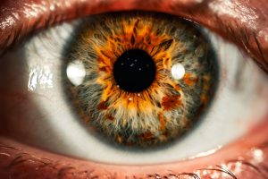


Vision
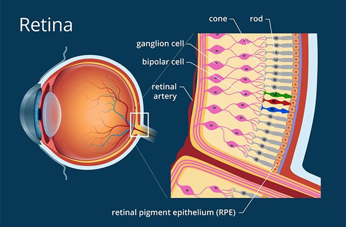
Vision starts with the eyes. The neural retinas in the eyes are part of the central nervous system, making the eyes the only part of the brain that sits outside the cranial vault. Three layers of neurons compose what we call the neural retinas. The job of the eyes is to collect light information and send it off to the rest of the brain, in a form that the brain can understand. No lights get past those neural retinas, but instead get received by the cells called photoreceptors (red, blue, and green cones). Light is received by the Retinal Ganglion Cells, then passed through the Bipolar-Amacrine-Horizontal Cells, and finally Rod and Cone Cells. There are two different types of photoreceptor cells called Rods and Cones. Cone cells are mainly responsible for
daytime vision and the Rod cells are mainly responsible for nighttime vision under low-light conditions. The Retinal Ganglion Cells (RGCs) receive this light and then send this information gathered to the rest of the brain. The cones in your eyes respond to the wavelengths of light that is reflected from an object, and these wavelengths are perceived by the brain for example as green color or round object. The brain compares the amount of green reflection coming off that object to the amount of red and blue around it. So, in theory we don’t see anything directly but instead the brain is comparing electrical signals to make sense of the objects it focuses on. The eyes basically are taking in light and this light is translated into electrical signals that the brain interprets.
AAV Media, LLC. © 2000-2023 AAV Media, LLC.
Depth Perception
The brain is creating a sense of depth, even though what arrives at the retina is in simple terms a 2-dimensional flat image, but how does the brain know what depth is. Things that are closer to you tend to be larger than things that are far away. Things that are closer to you tend to look like they are moving faster, for example if you are in a moving train at the sides things are moving very fast but in the distance things seem to move very slowly. The brain uses about 40-50% of its capacity for vision.
Evolution of The Eye
Eyesight did not evolve for us to see shapes, colors, motion, and form. The most ancient cells in our eyes, and the reason we have eyes, is to communicate information about the time of day to the rest of the brain and body. There are no extraocular photoreceptors meaning that it is impossible for light to get into all cells of the body, but every cell in the body needs to know if it’s day or night. There are so called melanopsin retinal ganglion cells named after the opsin that they contain within them. These melanopsin cells have their own photoreceptor built inside them. The opsin that they contain is very similar to the melanopsin that is present in the skin of some amphibians and that causes those amphibians to change their skin color in different light conditions. These RGCs communicate to areas of the brain when qualities of light are present in your environment, and signal to the brain that is early day or late in the day. These cells regulate when you’ll be feeling like sleeping, when you feel awake, how fast your metabolism is, your blood sugar levels, your dopamine levels, and your pain threshold. These melanopsin ganglion cells have been shown by many university neuroscience labs that they are essential to watch early morning and late afternoon rays of sun which will trigger your circadian clock that sits in the roof of the mouth, and this will trigger every cell in the body.
Visual Focus
The eye dynamically can adjust where light lands by moving the lens and changing the shape of the lens in your eye through a process called accommodation. Accommodation is our ability to accommodate things that are closer or further away from us. The Iris, musculature and the Ciliary body move the lens. When looking far away your lens relaxes flattening out but when it sees something up close it takes effort to focus on it. The Lens gets thicker to bring the light to the retina and not to a location in front of it or behind it. There are also changes in the size of the pupil as things get closer and further away from you. Healthy pupils are going to dilate when you look at something far away from you, but this also happens when you see something that excites or stresses you.
Vision ‘Eye to Brain Pathway’
Eye-to-Brain connection, also called the retinofugal pathway, is the most used sense human use to go about moving around the world. This pathway consists of the axons from the retinal ganglion cells (RCGs) which are the output neurons of the eye (Huberman). The eyes connect to the brain through the optic nerves. The optic nerves are a pair of nerves that transmit visual information from the eyes to the brain.
When light enters the eye, it passes through the cornea, the lens, and the vitreous humor (a gel-like substance that fills the eyes). The light is then focused on the retina, a layer of cells located at the back of the eye. The retina contains photoreceptor cells called rods and cones, which are responsible for detecting light and transmitting visual signals to the brain. The rods are sensitive to low light levels and responsible for night vision, while the cones are responsible for color vision and visual acuity. The information from the rods and cones is processed by other cells in the retina, and the resulting signals are then transmitted to the brain through the optic nerves. The optic nerves exit the back of the eye and travel to the brain, where they connect to the visual cortex, a region in the back of the brain responsible for processing visual information. Once the visual signals reach the visual cortex, they are further processed and interpreted to give rise to our perception of the visual world.
The visual interpretation of the brain involves multiple areas of the brain, including the primary visual cortex, secondary visual cortex, and higher-order visual processing areas. Here are the main parts of the brain involved in visual interpretation:
- Primary Visual Cortex (V1): Located in the occipital lobe at the back of the brain, the primary visual cortex is responsible for processing basic visual features such as edges, lines, and orientations.
- Secondary Visual Cortex (V2,V3,V4): These areas, located adjacent to V1, are responsible for more complex processing of visual information such as object recognition, color processing, and motion perception.
- Inferior Temporal Cortex (IT): Located in the temporal lobe, this area is involved in object recognition, color processing, and motion perception.
- Posterior Parietal Cortex (PPC): Located in the parietal lobe, this area is involved in spatial processing and perception, as well as the integration of visual information with other sensory modalities such as touch.
- Frontal Eye Fields (FEF): Located in the frontal lobe, this area is involved in the control of eye movements and attentional processes during visual perception.
- Fusiform Gyrus: A region located in the temporal lobe that is involved in face recognition and processing of the complex visual stimuli, such as words and numbers.
Eye health protocols
In the age of technology and smartphones, a lot of us spend a good amount of time looking at our phones or laptops or papers during our daily lives, and this is an unprecedented event in human history. For instance, myopia has increased exponentially across the world and it is increasing particularly faster in children and young adults who are looking at things at very close range. This is not to say that looking at things up close is bad for you, but it is too be considered to take measures if you want to keep good eye-health. The structure of the eye is such that the lens of the eye can move but also the length the eyeball from front to back is impacted by how close or how far you view objects during the day and especially during development. It is recommended that you get at least an hour of exposure to looking at this in the distance, and this could be done by going outside of the house and looking at things at a distance. This could be accomplished by running, hiking, walking, cycling or anything that requires you to look for things at distance. Another way to keep you vision healthy is to keep the musculature that controls the eyes movement and the change of shape of the lens when looking at things close to looking at things far away from you. This exercise is called Smooth-Pursuit Task, this exercise involves tracking a small object in front of you (example: pen, or dot) while this small object moves slowly and your focus follows this object. By doing this task for 2-3 minutes each day, you’re maintaining the ability of the musculature of your eye to do smooth pursuit. Another exercise is the Near-Far exercise, and this is properly done by holding an object straight in-front of your eyes (example: a pen or stick) and focusing on the tip of the object and then moving that object closer to you face until you can no longer have a crisp image of the objects tip. You might want to try this for a minute or two and try to play around that crisp-image threshold for a bit and then go back at full length. This might cause a bit if fatigue or strain in the eye but this is actually building up the musculature and neuromuscular connections around your eyes.
Exercises
Here are some exercises that can help improve eye health:
- Eye relaxation exercises: Close your eyes and breathe deeply for a few minutes, allowing your mind and eyes to relax. Then, open your eyes and focus on a distant object for 10-15 seconds, and then shift your focus to a nearby object for 10-15 seconds. Repeat this exercise several times.
- Eye blinking: Blinking helps to moisten and lubricate the eyes, which can reduce the risk of dry eyes and eye strain. Try to blink your eyes rapidly for a few seconds every 10-15 minutes while doing visually demanding tasks, such as reading or using a computer.
- Eye stretching exercises: Close your eyes and gently massage your eyelids with your fingers for 1-2 minutes. Then, look up and down, and then left and right without moving your head. Repeat this exercise several times.
Hearing
Mechanical function and Interpreting sound
Lets talk about hearing and how we convert sound-waves into meaning in our brains. The technical names for our ears are Pinnas, and these anatomical structures are made out of cartilage and skin tissue. Our ears are arranged in such a way that they can capture many different frequencies of sound. The shape of the ears are such that they amplify high and low frequency sounds, and these sound waves travel into our ears and reach the eardrums. When sound reaches the eardrum it starts to vibrate, and attached to the eardrum are three tiny bones called the malleus, incus and stapes. These three bones are attached to each other and form a hammer-like structure. The malleus is attached to the eardrum and the stapes is attached to an internal structure called the Cochlea. When the stapes starts vibrating, it vibrates or hammers into a coiled, fluid filled piece of tissue called the Cochlea. Inside the Cochlea is where sound is converted into electrical signals that the brain can interpret. One end the Cochlea is wider and more rigid than the other end so sound enters easily and is funneled down to a more narrow area with hairlike cells called cilia. These cells are very important in distinguishing between sounds that are low frequency and high frequency.
The process of distinguishing between low and high frequency is done within the coil of the Cochlea with the help of these inner hairlike cells. When these hairlike cells move, they send signals to the brain helping us distinguish where and what a sound is in our surroundings. The base of the Cochlea is very rigid and if the hair cells in this area move they will detect high frequency sounds. The top of Cochlea called the apex which is very flexible, and if their hair cells move they will detect low frequency sounds. It is important to note that there are high and low frequencies, but also in-between frequencies similar to the light-spectrum of colors. The Cochlea has the ability to distinguish between all variations of sound frequency.
The hair cells in the Cochlea transmit their movement patterns into the brain through axons, and this information arrives at different parts of the brain to make you conscious of where the sounds are in your environment. The Cochlea sends information to the spiral ganglion, which is a clump of neurons, and then the information travels further to the cochlear nuclei located in the brainstem and then is sent to the superior olive. Then the neurons from the superior olive send the information to the inferior colliculus, then to the medial geniculate nucleus and then finally up to the Auditory cortex where you make sense of that information. The function of the cochlea and the shape of our ears are vital to our survival by letting us know where and what a sound is in relation to our space in the environment. 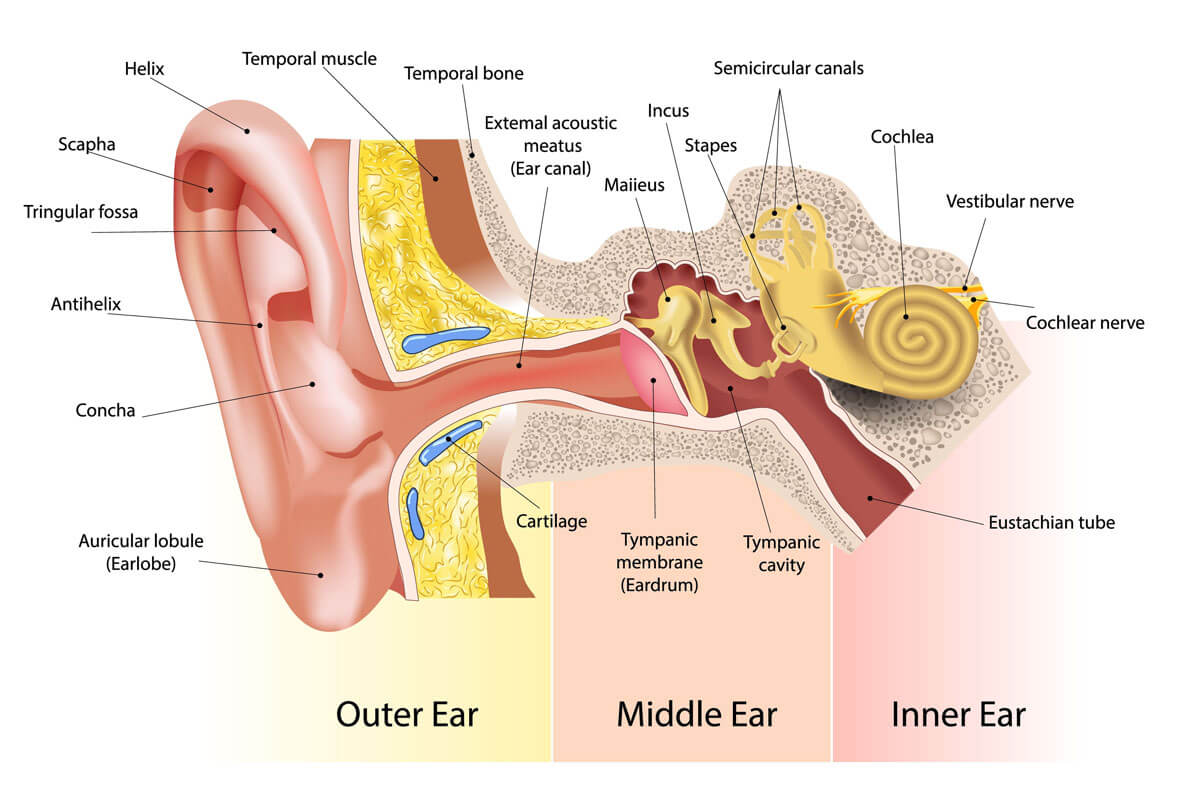
Happy Ears Hearing Center, LLC ® © 2014 – 2023 | All Rights Reserved
Localizing Sound
It’s vital for our survival to know if something is coming at us from the side, or if something is going to fall on top of us. Our ears aid our eyes in locating potential threats or obstacles. Our visual and auditory systems collaborate to help locate the position of objects in our surroundings. Deep in our brainstem, there is a station that is responsible for helping you identify where sounds are coming from through a process called inter-aural time differences. This process works by identifying sounds that land in one ear before the other, and these stations (neurons) in our brain calculate the difference in the time of arrival for those sounds waves between your left and right ear. If sound arrives at the same time in both ears, your brain assumes the sound comes from in front of you. If sound arrives first from you right then it comes from the right, and if it arrives first from you left then it comes from the left. The shape of your ears can modify the sound coming from below, in-front or above you making the ear very efficient on sound localization from different planes.
Here are some ways to improve hearing:
- Protect your ears: Exposure to loud noises over an extended period of time can cause hearing loss. Wearing earplugs or earmuffs in noisy environments and turning down the volume on personal music devices can help protect your ears.
- Quit smoking: Smoking has been linked to hearing loss, so quitting smoking can help improve your hearing health.
- Manage medical conditions: Certain medical conditions, such as diabetes and high blood pressure, can contribute to hearing loss. Managing these conditions through medication, diet, and exercise can help improve hearing health.
- Exercise regularly: Exercise has been shown to improve blood flow and oxygen supply to the ears, which can help improve hearing.
- Eat a healthy diet: A healthy diet that includes plenty of fruits, vegetables, and lean protein can help improve hearing health by providing essential vitamins and minerals.
- Get regular hearing tests: Regular hearing tests can help detect hearing loss early and allow for early intervention, which can help prevent further damage.
- Consider hearing aids: If you have hearing loss, hearing aids can help improve your ability to hear and communicate with others.
Overall, taking steps to protect your ears, managing medical conditions, and living a healthy lifestyle can help improve hearing health and prevent hearing loss.
Exercises
- There are several exercises you can do to help improve the health of your ears:
- Jaw exercises: Stretching and strengthening the muscles around the jaw can help to improve circulation to the ears and reduce tension. For example, try opening your mouth wide and holding the position for a few seconds, then slowly closing your mouth.
- Ear massages: Massaging the ears can help to stimulate blood flow and lymphatic drainage, which can help to reduce inflammation and improve hearing. Gently massage the earlobes and the area behind the ears with your fingers.
- Yoga and meditation: Practicing yoga and meditation can help to reduce stress and improve circulation, which can benefit ear health. Specific yoga poses, such as the shoulder stand or fish pose, can also help to improve circulation to the head and ears.
- Listening exercises: Exposing your ears to different sounds and frequencies can help to improve hearing. For example, try listening to music or sounds that you normally wouldn’t, or practice focusing on a particular sound in a noisy environment.
- Ear training exercises: These exercises involve training your brain to process sounds more effectively, which can improve hearing. For example, try listening to a speech recording at a low volume and gradually increasing the volume until you can hear it clearly.
Effects of aging on our hearing
Hearing loss affects about one-third of adults ages 61 to 70 and over 80 percent of adults over the age of 85. While certain medications, viruses, and diseases can cause hearing loss, it is mostly a genetic condition. The deterioration of cells in the inner ear is called presbycusis and is often the cause of hearing loss in elderly populations (Walling et al., 2012). One consideration that is becoming increasingly relevant to hearing loss is exposure to loud noises. Social events, concerts, and especially loud music in headphones are all very common in today’s society. Any prolonged exposure to loud noises such as these leads to excess deterioration of the inner ear and therefore one’s ability to hear worsens.
Communication is one of the most vital components of everyday life and maintaining relationships. Those afflicted with hearing loss at varying degrees of severity will have trouble communicating with others. This can be a particularly frustrating experience for all those involved. Not only that, poor hearing can worsen overall cognitive functioning in elderly populations as it takes them longer to process and understand information (Wingfield et al., 2005) Unfortunately, Americans must spend between $1,000 and $5,000 for one pair of prescription strength hearing aids, and often this cost is not covered by insurance (Jilla et al., 2020). This hefty price is worth it for some, but completely out of the question for others.
Current medical costs for the treatment of hearing loss place a large economic burden on elderly populations. It’s important to recognize that there are methods of preventing hearing loss before it starts. While most hearing loss is a natural part of the aging process, loud noises over time, a poor diet, and taking ototoxic medications can lead to the process speeding up and worsening (Fausti et al., 2005). Eating a diet rich in omega-3 fatty acids can lower the severity and likelihood of hearing loss as well. Wearing ear plugs in loud environments and limiting excessively loud music from speakers or headphones helps to protect the delicate structures of the inner ear as well.
Touch
SKIN
Human skin is the largest organ in the body human covering around 2 meters2 of area, and service as the first layer of physical defense against the outside environment. The skin serves a plethora of functions such as thermoregulation, tactile sensory perception, physical protection, metabolic and biochemical functions, and immunity functions (Wysocki, A. B., 1999).
Skin anatomy
The skin has many three mayor layers and sublayers, and they all serve a specific function. These layers are, starting with the outermost layer, the Epidermis (Stratum Corneum, Stratum Lucidum, Stratum Granulosum, Stratum Spinosum, and Stratum Basale.), Basement Membrane, the Dermis, and the Hypodermis/ Superficial Fascia. The Epidermis (75 to 150 µm and up to 0.6 mm) hold five sublayers that each of them have a specific function like the S. Corneum is composed of dead skin cells and contains keratin (filaments proteins) and its resistant to changes in pH, temperatures, enzymatic digestion. The S. Lucidum is an area of intense enzymatic activity, the S. Granulosum is packing electron-dense basophilic staining granules (keratohyalin granules) aiding in assembly of keratin filaments, the S. Spinosum where synthesis of involucrin protein forming cornified envelopes, and the innermost epidermal layer S. Basale which is the layer where cells are going into mitosis (Wysocki, A. B., 1999). Going deeper into the skin is the Basement Membrane serving as the layer between the Epidermis and Dermis by regulating the transfer of proteins and other materials, the Dermis (2 to 4 mm) serves as the anchor of the skin layers acting as webs and also restore skin integrity just to name a few features, and finally the Hypodermis/Superficial Fascia supports skin structure and contains blood vessels and nerve endings.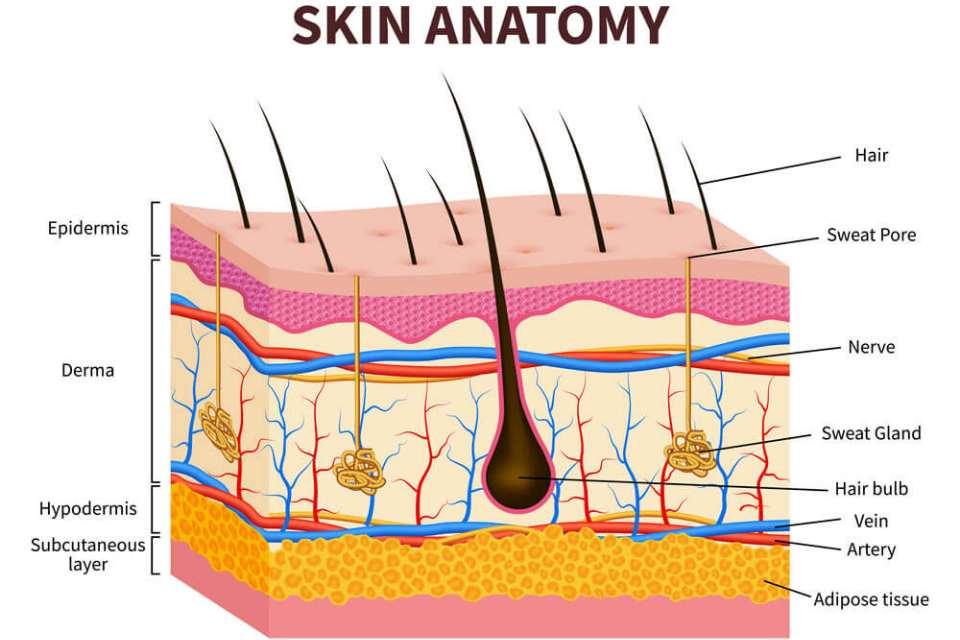
© 2023 The Dermatology Specialists. All Rights Reserved.
Sensory Function of Skin
The skin senses touch through specialized nerve endings called mechanoreceptors. These mechanoreceptors are located in the skin and detect mechanical pressure, vibration, stretch, and other physical forces(Wysocki, A. B., 1999). There are several types of mechanoreceptors in the skin, each of which responds to a specific type of touch stimulus. The two most common types of mechanoreceptors are Meissner’s corpuscles and Merkel cells. Meissner’s corpuscles are located close to the skin surface and are particularly sensitive to light touch and vibration, while Merkel cells are located deeper in the skin and respond to sustained pressure and texture. When a stimulus, such as pressure or vibration, is applied to the skin, it causes the mechanoreceptors to be deformed or stretched. This deformation generates an electrical signal that is sent to the brain via nerve fibers. The brain then interprets these signals to determine the location, intensity, and type of touch stimulus that has been applied to the skin. Overall, the skin’s ability to sense touch is a complex process that involves a combination of specialized nerve endings, nerve fibers, and the brain’s interpretation of these signals.
Here are some key takeaways about the function of the skin:
- Protection: The skin serves as a barrier between the body and the external environment, protecting the body from harmful agents such as bacteria, viruses, and physical trauma.
- Sensation: The skin is richly innervated with nerve endings that allow us to sense touch, temperature, pressure, and pain.
- Thermoregulation: The skin plays an important role in regulating body temperature through the dilation or constriction of blood vessels and the production of sweat.
- Vitamin D synthesis: The skin is capable of synthesizing vitamin D from sunlight, which is important for bone health and immune function.
- Absorption: The skin can absorb certain substances, such as topical medications or toxins, that come into contact with it.
- Excretion: The skin can also excrete certain substances, such as water and salt, through sweating.
Exercises
Here are some exercises and lifestyle changes that can help improve skin health:
- Exercise regularly: Exercise increases blood flow and oxygen to the skin, which can help nourish skin cells and improve skin tone.
- Stay hydrated: Drinking plenty of water helps to keep the skin hydrated and can improve the appearance of fine lines and wrinkles.
- Eat a healthy diet: A diet rich in fruits, vegetables, and healthy fats can provide the nutrients necessary for healthy skin, such as vitamins A, C, and E, as well as antioxidants.
- Practice stress management: Stress can contribute to skin problems such as acne and eczema, so practicing stress-management techniques such as meditation, yoga, or deep breathing can help improve skin health.
- Get enough sleep: Sleep is essential for healthy skin, as it allows skin cells to regenerate and repair. Aim for at least 7-8 hours of sleep per night.
- Practice good skincare: This includes cleansing the skin regularly, using a moisturizer, and protecting the skin from the sun with sunscreen.
- Facial exercises: These exercises can help improve blood circulation to the face and stimulate collagen production, which can improve the appearance of the skin. Examples include facial massages, facial yoga, and facial acupressure.
Please provide your feedback here
Barnes, G. R. (2008). Cognitive processes involved in smooth pursuit eye movements. Brain and cognition, 68(3), 309-326.
Fausti, S. A., Wilmington, D. J., Helt, P. V., Helt, W. J., & Konrad-Martin, D. (2005). Hearing
health and care: The need for improved hearing loss prevention and hearing conservation practices. The Journal of Rehabilitation Research and Development, 42(4s), 45. https://doi.org/10.1682/jrrd.2005.02.0039
Jilla, A. M., Johnson, C. E., & Huntington-Klein, N. (2020). Hearing aid affordability in the
United States. Disability and Rehabilitation: Assistive Technology, 18(3), 246–252. https://doi.org/10.1080/17483107.2020.1822449
Litzinger, T. C., & Del Rio-Tsonis, K. (2002). Eye anatomy. Encyclopedia of life science.
Photo of Eye by Colin Lloyd on Unsplash (2021)
Photo of Soundwave by Pawel Czerwinski on Unsplash (2021)
Photo of Hand by Alexei Scutari on Unsplash (2018)
Risoud, M., Hanson, J. N., Gauvrit, F., Renard, C., Lemesre, P. E., Bonne, N. X., & Vincent, C. (2018). Sound source localization. European annals of otorhinolaryngology, head and neck diseases, 135(4), 259-264.
Sundar, P. S., Chowdhury, C., & Kamarthi, S. (2021). Evaluation of human ear anatomy and functionality by axiomatic design. Biomimetics, 6(2), 31.
Walling, A. D., & Dickson, G. M. (2012). Hearing Loss in Older Adults. American
Family Physician, 85(12), 1150–1156. https://www.aafp.org/pubs/afp/issues/2012/0615/p1150.html
Wingfield, A., Tun, P. A., & McCoy, S. L. (2005). Hearing Loss in Older Adulthood. Current
Directions in Psychological Science, 14(3), 144–148. https://doi.org/10.1111/j.0963-7214.2005.00356.x
Wysocki, A. B. (1999). Skin anatomy, physiology, and pathophysiology. Nursing Clinics of North America, 34(4), 777-797.