3 Cerebellum
pr379
To whomever may be reading this little snippet. My name is Peter, and I am an armchair cerebellum enthusiast. Being a Masters student in Kinesiology, I have had a number of classes explaining the basic functions and location of the cerebellum, but never much more than a baseline understanding. I have always been fascinated by it however, as it is known that the cerebellum plays a huge role in information processing and learning motor patterns. I am fascinated by this idea, as I heavily rely on my body to perform many of the things I love and hold dear: playing music (piano, guitar, etc.), lifting weights, and doing just about anything I can outdoors. Something like learning a piece on the piano relies heavily on learning and repeating movement patterns. Control for minute details down to the amount of force each finger applies to the keys giving them soft or powerful tones, the speed of the force applied resulting in a sustained or staccato notes, and of course stringing together some insanely intricate, interstellar-esque madness are all heavily reliant on the cerebellum. This chapter will explore some of the anatomy and processing involved with the cerebellum, as well as what damage and disease to the cerebellum can do to quality of life. We will also discuss how to observe activity in the cerebellum, and what future research may look like in this area of neuromechanics. There is some funny, confusing terminology in here, so bear with me. If you hope to add, revise, or correct any of my work, I only ask that you find a way to let me know, so I may have a better understanding of this totally cool structure that may or may not have inspired one of my tattoos (just wait until you see what a purkinje cell looks like).
Happy reading!
Introduction
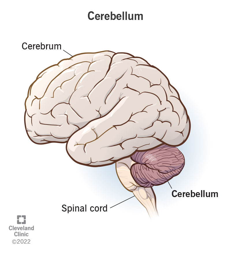
https://my.clevelandclinic.org/-/scassets/images/org/health/articles/23418-cerebellum
The Cerebellum, also known as the “hindbrain,” or “little brain,” occupies the posterior portion of the cranium, inferior to the occipital and temporal lobes. While all of the contributions to brain function are not fully understood, it is clear that the cerebellum plays an important role in motor coordination, motor learning, and predicting and perfecting movement patterns. Think learning to walk at a young age, or perhaps rolling a coin across your knuckles. Every micro-movement, amount and direction of force production, body position, reaction time, and body awareness, or proprioception, is integral to performing the task effectively. If any component is performed inefficiently, the cerebellum contributes to learning, relearning, and correcting these micro-patterns.
It is commonly described that the cerebellum helps learn specific motor patterns, and the motor cortex hits the save button on said patterns. This is how someone who may no longer remember how to play the piano will still be able to bust out some hot cross buns or fur elise, even if it has been years since they practiced. The development of learned movement and thought patterns can be described as neuroplasticity, as new neural pathways are developed and strengthened with repeated reinforcement. For more on neuroplasticity, see chapter six.
Optimal cerebellar function will aid in both online and offline learning, as well as error detection and correction. While most would assume that the cerebellum would most heavily contribute to online learning, which is in essence someone learning learning a given movement real time with both feedforward and feedback response (these will be discussed in greater detail below), Samaei et al. 2017 has shown that transcranial stimulation of the cerebellum may improve offline learning (learning in the absence of movement practice) as well. For example, someone practicing shooting a free throw, and correcting for detected errors is developing online learning capacity using a combination of feedforward and feedback responses. Of course, a variety of factors would influence performance including stress, attention span, energy level, degree of skill development, learning environment, wakefulness, and so forth. However, all offline learning this individual goes through would take place separate from the physical practice of the free throw itself through repetitive thought and rumination of said activity.
Like any other system in the brain, the affinity for growth, activity, and development of cerebellar function varies from one individual to another. This can be observed among those assholes who just seem to pick up a skill with seemingly no “learning curve,” or practice, while others may need to take a much more meticulous approach if they wish to perfect a skill. There is plenty of evidence however that the longevity and overall function and health of the cerebellum and surrounding structures can be enhanced if prioritized and challenged at younger ages of development. This can be observed among children who grow up bilingual throughout their developmental stages, becoming fluent at a much faster rate than an adult learning a new language (De Smet et al., 2013). Not only that, but these bilingual children are more likely to retain their language skills later in life as well, even if they have gone without practice for years. This is logically sound, with neuroplasticity being considered to be more fertile in younger stages of development (Kolb & Gibb, 2011).
As we will discuss, there is recent mounting evidence that the cerebellum plays a much larger role with higher executive function involving cognition, emotion, memory, and more. This only furthers the functional connectivity between the cerebellum and other structures taking up both nearby real estate within the brain and surprisingly far.
Anatomy
Placed dorsal to the brain stem and inferior to the temporal and occipital lobes, the cerebellum occupies only 10% of the brain’s total volume, weighing on average 130 grams. However, this hunk of meat is estimated to contain between 50-80% of all neurons located in the brain (Knierim, 2022). In case you didn’t already know, the brain has an estimated 86 billion neurons, allowing the cerebellum between 43 and 69 billion to work with. This surplus of neural activity aids extensively to micro-calculations and adjustments to movement patterns and faster learning (Mirdamadi & Block, 2021).
The Cerebellum is attached to the brainstem and transmits input and output signaling from the 3 paired cerebellar peduncles (inferior, middle, superior). Output from the cerebellum is regulated solely by the Deep Cerebellar Nuclei, which is encased by dense, neurally dense, folded gray matter tissue called the cerebellar cortex (Knierim, 2022).
The cerebellum can be split into two different hemispheres, divided by the Vermis. Within these hemispheres are different functional zones (Knierim, 2022).
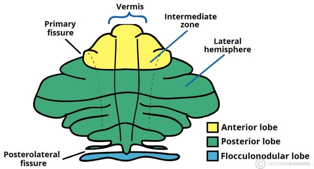
https://teachmeanatomy.info/neuroanatomy/structures/cerebellum/
| Lateral Zone | spinocerebellum | Muscle tone regulation | Carries info from:
Muscle spindles GTO Joint Receptors |
| Intermediate Zone | cerebrocerebellum | Assisting, planning, programming fine motor movements | Carries info from:
Muscle spindles GTO Joint receptors Trunk Lower extremities |
| Median/Vermal Zone | vestibulocerebellum | Posture and balance |
The lateral zone is as its name implies on the outermost part of both hemispheres, and aids in planning and programming movements by communicating with the dentate nuclei. The Intermediate Zone is medial to the lateral zone (so in between the Lateral Zone and Vermis), and likes to communicate with the Interposed nuclei. Finally, there is the Median/Vermal Zone, which communicates with the fastigial nuclei (Knierim, 2022). I should mention that as I understand it, the dentate, interposed, and fastigial nuclei all fall under the umbrella term of “deep cerebellar nuclei.”
There are 3 major lobes within the cerebellum: Anterior, Posterior, and Flocculonodular. The Anterior and Posterior Lobes are separated by the primary fissure, while the posterior and flocculonodular lobes are separated by the posterolateral fissure. (Cerebellum, 2022)
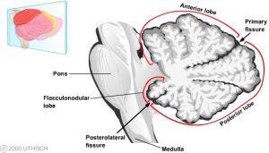
https://nba.uth.tmc.edu/neuroscience/m/s3/chapter05.html
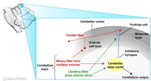
Essentially, input will pass through the peduncles, to the mossy fibers. The mossy fibers will then transmit the signal to the granule cells, with one fiber innervating hundreds of granule cells (Cerebellum, 2022). Granule cells are densely packed neurons that make up the innermost layer of the cerebellar cortex, interwoven with dense, complex dendritic neurons called purkinje cells (Knierim, 2022). The relationship between the purkinje cells and the deep cerebellar nuclei is complex and quite frankly very confusing, but my best understanding is that the purkinje cells solely regulate output of the cerebellar cortex to the deep cerebellar nuclei, and the deep cerebellar nuclei regulate output from the entirety of the cerebellum. These purkinje cells are innervated by both climbing fibers that originate from the inferior olive, and input from the previously mentioned mossy fibers (Knierim, 2022). Purkinje cells also receive inhibitory signals from parallel fibers that run horizontally in the molecular (outermost) layer of the cerebellar cortex ( Knierim, 2022).
Piersol, George A. Human Anatomy (Philadelphia, PA: J. B. Lippincott Company, 1908)
Processing
Afferent (incoming from sensory neurons) and Efferent (outgoing from brain) signals are constantly being sent to and from the cerebellum via the peripheral and central nervous systems to carry out tasks (Cerebellum, 2022). Visuomotor stimulus and Proprioceptors throughout the body provide input through the middle and inferior peduncles, while all output signals are transmitted through the Deep Cerebellar Nuclei, and sequentially the inferior and superior peduncles (Knierim, 2022). To minimize the confusion (a lot of big words going on) I will include a graphic of how input and output flows through the cerebellum.
https://nba.uth.tmc.edu/neuroscience/m/s3/chapter05.html
There also appears to be a distinct topographical difference in where emotion and cognition are regulated. I have a limited understanding, but it sounds like the sensorimotor cerebellum is regulated by the anterior lobe, as well as lobules VI and VIII (Stoodley, 2010). Cognition within the cerebellum seems to be influenced by activity primarily in lobules VII and VI, as well as the ventral dentate nucleus. Finally, the fastigial nucleus and posterior nucleus make up the topographical real estate representing the limbic cerebellum, or the portion of the cerebellum dedicated towards sensory processing (Stoodley, 2010). Of course, you cannot have sensorimotor without sensory processing, so you can imagine how like many other structures in the brain, there is a significant amount of cohesion and fluidity between regions, as opposed to rigid roles.
Feedback Control Systems:
Feedback control systems are as they sound. When something other than the desired outcome occurs, the cerebellum plays a role in adjusting for any discrepancies or over/underestimations (Cerebellum, 2022). Once again, we can use learning to walk as an example, with a toddler continuing to make adjustments to balance, force production, angle of their joints, etc. This is a slow process, and requires “failure,” in order to make alterations, but is an incredible learning tool to develop and maximize efficiency of complex movement patterns (Mirdamadi & Block, 2021).
As I understand it, both feedback and feedforward errors that are detected and corrected are primarily regulated by complex signals sent by the climbing fibers of the inferior olive to the purkinje cells (Knierim, 2022). These purkinje cells are integral in regulating and correcting for errors both detected via motor and sensory pathways, and will directly influence the output sent by the cerebellar nuclei. Interestingly, purkinje cells in different physical locations within the cerebellar cortex will be activated depending on type of sensory error detected (motor, visual, auditory, etc) and whether or not the error was via feedback or feedforward detection (Stoodley, 2010). When a successful movement pattern is finally achieved, there is a measurable (via fMRI) spike in the corresponding nuclei depending on type of stimulus (Ito, 2013).
Feedforward Control Systems:
Here is the fun one. It has been suggested that the cerebellum can also use the overload of sensory information provided to predict outcomes, and alter movements to yield a positive effect (Knierim, 2022). This allows us as humans to be more proactive, and act before receiving input from a feedback system. We are able to make adjustments and alterations to a given pattern without having to wait for feedback, and instead use our diverse range of sensory information to predict and readjust. The Vestibulo-ocular Reflex (VOR) if you haven’t heard of it, is this really cool compensatory movement of the eyes that automatically adjusts for movement of the head to maintain smooth, continual vision, and presents a wonderful example of feedforward control (Cerebellum, 2022; Knierim, 2022). For more information on VOR, check out chapter 7. One other reflex titled the saccadic eye movement is essentially this ballistic eye movement in an attempt to follow a target that is moving too fast for visual feedback (Kheradmand, 2011). This allows us to create a projected end-point with no feedback or tracking required, allowing for predicted “reactions,” before any actual result has occurred. This being said, there is a degree of trial and error involved. Any skill we spend time developing takes significant time and effort, so predicting outcomes will be more effective if it is a familiar movement pattern (Kheradmand, 2011). Kicking a moving soccer ball will use a combination of feedback and feedforward processes, and requires numerous attempts and reattempts to coordinate the corresponding muscle groups to contract and stretch reliably, and is influenced heavily by speed of the ball, speed of the individual, the force required to kick, how wet the grass is, etc. Therefore, a seasoned athlete with years of experience will be able to more effectively make use of this feedforward system and predict a soccer ball’s positioning compared to a novice in the sport (Kolb & Gibb, 2011).
Disorders
So there are a number of conditions that can result from physical damage to the cerebellum. However, many are not exclusive to the cerebellum. Like many structures in the brain, processes and functions are not specific to the “one and only,” this or that. Rather, many of these structures work in tandem to provide fluid and dynamic regulation of body processes. Symptoms such as ataxia, or an inability to control voluntary movements can be influenced by cerebellar damage, damage to the vestibular system or simply loss of proprioception due to neuropathy or some other loss of sensory input (Cerebellum, 2022). Lesions in the cerebellar hemispheres can cause both hypometria and hypermetria, which is essentially undershooting and overshooting coordinated voluntary movements, respectively (Knierim, 2022). Damage to the cerebellum can also cause conditions like dysarthria and scanning speech, which will impair speech patterns and jack up the body’s ability to effectively control the tongue and lips. Observable symptoms include slowed speech, inconsistent pauses, mumbling and changes in pitch and speed (Cerebellum, 2022).
There are a wide variety of alarmingly specific conditions both directly and indirectly influenced by impaired cerebellar function relating to speech, vision, motor control, etc. There are also a number of in-depth ataxia classifications both congenital and acquired. I will not list all of them as I feel it is a tad redundant and they have insane names like dysdiadochokinesia, but boiled down, corrupted cerebellar structure or function, as well as connectivity with surrounding structures such as the basal ganglia can really impair motor control, emotional regulation, learning patterns and cognitive function (Cerebellum, 2022; Samaei et al, 2017; Knierim, 2022). I will discuss this in more detail in the next section, but new research is supporting claims that underactive cerebellar function is associated with a number of non-motor specific disorders including autism, schizophrenia, Parkinson’s disease, substance use and post traumatic stress disorders (De Smet, 2013; Grimaldi et al, 2016; Mirdamadi, 2016).
Current/Future research
Activity in the cerebellum can be potentially influenced or modulated using a technique called Transcranial Direct Current Stimulation or tDCS (Sameai et al, 2017). As my monkey brain understands it, Anodal (positive current) and Cathodal (negative current) electrodes are placed near the site of interest to manipulate how current is flowing to and from specific brain regions (Sameai et al., 2017). More recently, Cerebellar Transcranial Alternating Current Technique (cb-tACS) has been shown to be an effective electrophysiological technique for modulating both motor and cognitive functions (Wessel, 2023). I don’t really understand the difference between these two techniques other than one uses direct current and the other uses alternating current, but ultimately both have been suggested to be effective tools for “resetting,” cerebellar network activity (Samaei et al., 2017; Wessel, 2023). Continuous theta burst stimulation is another technique by which repetitive transcranial stimulation can alter cortical function and activity (Mirdamadi & Block, 2021). Of course, magnetic resonance imaging (MRI) is an effective tool for observing activity of the cerebellum and can contribute to our understanding of executive function, and to observe degree of functional connectivity (FC), which essentially describes one or more brain structures that will show similar activity or activation patterns in a variety of cognitive and emotional conditions (Habas, 2021). There are some limitations with using MRI, as there is no way to discern if connectivity between structures is correlational or causational.
While it is known that the cerebellum contributes heavily to motor control and movement patterns, there is more emerging evidence that it plays a larger role than previously understood in thought processes, memory storage, and emotional regulation (De Smet et al., 2013; Fastenrath et al., 2022). It has been suggested by a large-scale study performed out of the University of Basel that cerebellar activity plays a crucial role in storage of emotional memories and experiences (Fastenrath et al., 2022). While this mechanism can play an influential role in evolutionary development of humans, this may help explain how development of post-traumatic stress occurs, and perhaps better understand how to help treat it. Researchers out of the Albert Einstein College of Medicine in New York have suggested that the cerebellum is linked to the Ventral Tegmental Area (VTA) of the brain, which is heavily involved with motivation and reward behaviors. There were significant ties between cerebellar and VTA activation among mice reward habits, as well as social processing behaviors (Carta et al., 2017). This suggests that many disorders relating to reward and social and emotional behaviors may be linked to dysfunction of the cerebellum.
As I continue to read through studies posted since 2023, nearly all of them make the observations that the cerebellum plays a much greater role in non-motor related processes than previously thought, ranging from anxiety and psychiatric diseases to language control and emotional development.
Quiz Time!
ACTIVITY TIME
grab your prism goggles and hacky sacks, lets do this
Materials:
- plastic safety goggles
- two fresnel prisms for vision correction (30 diopter preferred)
- masking tape
- beanbags/hacky sacks
- scissors and pencil
Assembly:
- cut and lay the adhesive side of prisms to fit the goggles (ensure prism facets point in same direction on both sides)
- make a target with masking tape anywhere between 12-16 in in width about 3 m from your starting point
Procedure:
- throw 3-5 beanbags at the target, measuring the distance from the target each throw.
- put on the prims goggles and repeat the same number of throws (note: having a partner help retrieve and supply you with beanbags will simplify things)
- remove the goggles, and perform a final round of throws as quickly as possible
Analysis:
So what’s going on here? You may have noticed that applying the prism goggles resulted in an understandable immediate drop in accuracy compared to your first round of throws. If you had enough practice with the prism goggles, we would expect you to begin to hone in, and your accuracy would improve as you make the necessary adjustments to “rewrite,” the correct motor patterns that dictate success (i.e. hitting the target). However, upon removal of the prism goggles, you can expect a period of confusion with you newly recalibrated movement patterns being once again thrown for a loop. Try counting how many throws it takes you to regain the same degree accuracy as the primary round of throws.
Authors notes
Thanks for reading everybody! It is my understanding that many of you will be contributing to what I have gotten started for you. Once again, I would like to highlight that any alterations, clarifications, or corrections are absolutely welcome. I am by no means an expert, and I am sure that I made some incredibly broad claims, or perhaps miswrote some important details (I don’t think I did but who knows). I know I barely scratched the surface, but I hope I painted a well enough picture that helped build a well-rounded understanding of one of the coolest brain structures (they are all just so cool though, it’s hard to pick). I look forward to returning to this textbook in the future and seeing how you all improve and make this your own. Cheers!
Please provide your feedback here.
Citations
Cerebellum – Knowledge @ AMBOSS. (2022). https://www.amboss.com/us/knowledge/Cerebellum/#:~:text=Afferent%20cerebellar%20tracts%20(input),mossy%20fibers%20and%20climbing%20fibers
Cerebellum (Section 3, Chapter 5) Neuroscience Online: An Electronic Textbook for the Neurosciences | Department of Neurobiology and Anatomy – The University of Texas Medical School at Houston. (n.d.). https://nba.uth.tmc.edu/neuroscience/m/s3/chapter05.html
Ito M. Error detection and representation in the olivo-cerebellar system. Front Neural Circuits. 2013 Feb 22;7:1. https://doi.org/10.3389/fncir.2013.00001
What is the function of the middle cerebellar peduncles? a) they contain neurons that produce…
Carta, I., Chen, C. H., Schott, A., Dorizan, S., & Khodakhah, K. (in press). Cerebellar modulation of the reward circuitry and social behavior. Science. Online January 17, 2019. https://doi.org/10.1126/science.aav0581
Matthias Fastenrath, Klara Spalek, David Coynel, Eva Loos, Annette Milnik, Tobias Egli, Nathalie Schicktanz, Léonie Geissmann, Benno Roozendaal, Andreas Papassotiropoulos, Dominique J.-F. de Quervain. Human cerebellum and corticocerebellar connections involved in emotional memory enhancement. Proceedings of the National Academy of Sciences, 2022; 119 (41) DOI: 10.1073/pnas.2204900119
Wessel, M.J., Draaisma, L.R. & Hummel, F.C. Mini-review: Transcranial Alternating Current Stimulation and the Cerebellum. Cerebellum 22, 120–128 (2023). https://doi.org/10.1007/s12311-021-01362-4
Grimaldi G, Argyropoulos GP, Bastian A, Cortes M, Davis NJ, Edwards DJ, Ferrucci R, Fregni F, Galea JM, Hamada M, Manto M, Miall RC, Morales-Quezada L, Pope PA, Priori A, Rothwell J, Tomlinson SP, Celnik P. Cerebellar Transcranial Direct Current Stimulation (ctDCS): A Novel Approach to Understanding Cerebellar Function in Health and Disease. Neuroscientist. 2016 Feb;22(1):83-97. https://doi.org/10.1177/1073858414559409
Jasmine L. Mirdamadi, Hannah J. Block, Somatosensory versus cerebellar contributions to proprioceptive changes associated with motor skill learning: A theta burst stimulation study, Cortex, Volume 140, 2021, Pages 98-109, ISSN 0010-9452, https://doi.org/10.1016/j.cortex.2021.03.019.
Jasmine L. Mirdamadi (2016). Cerebellar role in Parkinson’s disease. Journal of neurophysiology. 116(3). https://doi.org/10.1152/jn.01132.2015
Samaei A, Ehsani F, Zoghi M, Hafez Yosephi M, Jaberzadeh S. Online and offline effects of cerebellar transcranial direct current stimulation on motor learning in healthy older adults: a randomized double-blind sham-controlled study. Eur J Neurosci. 2017 May;45(9):1177-1185. doi: 10.1111/ejn.13559
Hyo Jung De Smet, Philippe Paquier, Jo Verhoeven, Peter Mariën. (2013). The cerebellum: Its role in language and related cognitive and affective functions, Brain and Language, Volume 127, Issue 3, Pages 334-342. https://doi.org/10.1016/j.bandl.2012.11.001.
Kolb B, Gibb R. Brain plasticity and behaviour in the developing brain. J Can Acad Child Adolesc Psychiatry. 2011 Nov;20(4):265-76. PMID: 22114608; PMCID: PMC3222570.
Knierim, J. (2022, October 20). Cerebellum (section 3, Chapter 5) neuroscience online: An electronic textbook for the Neurosciences: Department of Neurobiology and Anatomy – the University of Texas Medical School at Houston. Cerebellum (Section 3, Chapter 5) Neuroscience Online: An Electronic Textbook for the Neurosciences | Department of Neurobiology and Anatomy – The University of Texas Medical School at Houston. Retrieved April 25, 2023, from https://nba.uth.tmc.edu/neuroscience/m/s3/chapter05.html
Cerebellum. https://radiologykey.com/cerebellum/. Updated: January 21, 2016. Accessed: April 26, 2023
Stoodley CJ, Schmahmann JD. Evidence for topographic organization in the cerebellum of motor control versus cognitive and affective processing. Cortex. 2010 Jul-Aug;46(7):831-44. https://doi.org/10.1016/j.cortex.2009.11.008
Kheradmand, A., & Zee, D. S. (2011, August 8). Cerebellum and ocular motor control. Frontiers. Retrieved April 26, 2023, from https://www.frontiersin.org/articles/10.3389/fneur.2011.00053/full https://doi.org/10.3389/fneur.2011.00053
Habas C. Functional Connectivity of the Cognitive Cerebellum. Front Syst Neurosci. 2021 Apr 8;15:642225. https://doi.org/10.3389/fnsys.2021.642225
dense, large-branching dendritic cells in charge of regulating information to the deep cerebellar nuclei. Acted upon by climbing fibers and mossy fibers
development of a skill or pattern in real time relying on a combination of sensory, feedback, and feedforward processes.
Development of a movement or pattern that is improved while not directly practicing said pattern.
one or more brain structures that show similar levels of activity in tandem in a given emotional or cognition pattern.
sole regulator of output of the cerebellum. Regulated in turn by the purkinje cells
Dense, folded gray matter encasing the deep cerebellar nuclei, comprised mainly of granule and purkinje cells
umbrella term describing an inability to control voluntary movements.